Menu
Meetings
Overview
About
Current Meeting
Last Meeting
Council & Foundation
History
Participations
Overview
Nobel Laureates
Young Scientists
Young Economists
Lindau Alumni
Guests
Partners & Support
Overview
Success Stories
Academic Partners
Benefactors & Contributors
Funding Opportunities
Responsibility
Outreach
Overview
Livestreams
Blog
News
Mission Education
Lindau Initiatives
Virtual Experience
Media
Mediatheque
Contact
Laureates
Meetings
Recordings
Topics
Educational
Lindau Mediatheque
Laureates
Meetings
Recordings
Topics
Educational
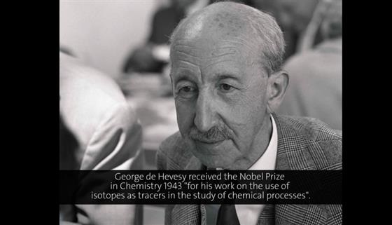
George de Hevesy