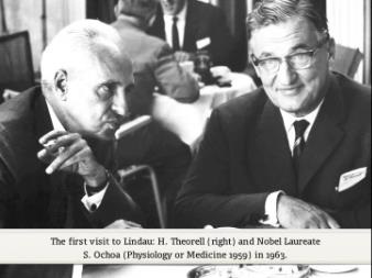
Hugo Theorell only gave two lectures at the Lindau Meetings. The first was in 1963 and its topic was how alcohol is burnt in the liver