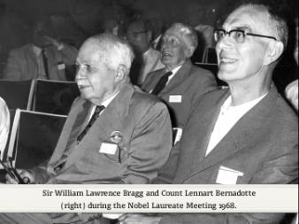
Count Bernadotte, Ladies and gentlemen.
I would first like to take the opportunity to thank you all and Count Bernadotte particularly
for the invitation which brings us here.
This is unfortunately the first time that I have been able to attend one of these occasions.
And I realise how much I have missed.
I would only like to point out to Count Bernadotte how widely my subject extends.
A few years ago the young people who were working in my physics laboratory,
two of them shared the Nobel Prize for chemistry and two of them shared the Nobel Prize for medicine.
There is therefore, Count Bernadotte, no reason that I can see why you should not invite me every year to...
That is of course if you like to do so.
A story I want to tell you is the history of a research which lasted over 25 years.
I have always been interested in trying to apply x-ray analysis to more and more complicated bodies.
And this story is the analysis of the most complicated bodies which have yet been successfully investigated,
the protein structures.
When Perutz and I started on this 25 years ago, success seemed almost impossible, they were so complicated.
But the prize, the reward for getting them out was a dazzling one.
And we felt we must have a try.
Now, perhaps I may just outline the problem.
The protein molecules are large molecules which play a part in the processes of life.
Haemoglobin, the one with which we started, is a molecule containing 10,000 atoms.
It has the special function in the body of conveying oxygen from the lungs to all parts of our body
and then taking back the carbon dioxide again to be given out in the lungs.
When we started this investigation, the haemoglobin which we chose is a molecule containing 10,000 atoms.
So far, the most complex structures which had been investigated were those done by Dorothy Hodgkin.
In particular the structure of vitamin B12, which contains 181 atoms.
The difficulty of getting out a structure goes up as a high power of the number of atoms in it.
So we were trying to do a molecule with 10,000 atoms when the best so far had 180.
May I remind you of the nature of x-ray analysis.
X-rays fall on a crystal structure composed of the molecules which you are examining.
You notice how strongly the x-rays are diffracted by the molecule in different directions.
And from this you have to deduce the arrangement of the molecules.
You make a measurement on diffraction and the result is the positions of the atoms.
Well, may I outline for you the nature of the problem in its full fearfulness.
The molecule contains 10,000 atoms, now we have to find their positions.
And their positions are defined by their coordinates, what we call parameters.
So as we have to find X, Y, Z, the coordinates of each atom, there are 30,000 variables in our equations,
Actually it is not as bad as that because fortunately, if I may draw on the board for a moment,
the molecule of haemoglobin has an axis of symmetry.
So if you know the position of an atom there, you know the position of its partner over there.
So we have 15,000 variables only to determine.
Now, we have enough equations to determine those variables.
Because each spot of the diffraction pattern gives us one equation.
A function for the first spot of 15,000 variables equals something we can measure,
the amplitude of the x-rays that make that spot.
And then you pass to the next spot, F2 and A2 and there are 30,000 of these equations.
So that then is the problem, to solve 30,000 equations, which are more than sufficient to determine 15,000 variables.
When we proposed to do this I went to Mellanby who was at that time the head of the medical research council
and I asked him for money, that is of course why one goes to people like Mellanby, and I said:
On the other hand, the importance of getting it out, if we are successful, is practically infinity.
And if you multiply zero by infinity it is possible that you will get something.”
And I’m very glad to say that he agreed and that started our research.
Now, what was my part in this research, because I think it is very wrong for heads of laboratories
to talk too much about the work which their young people do, it should be left to the young people to do that.
But I can perhaps again on the board illustrate what my part was, ...
going back to earth was WLB,
going into a protiate orbit was Perutz.
I think that perhaps describes it, that I was the first stage of the rocket which got this research off the ground...
What is the nature of a protein molecule?
It is very interesting the way in which nature builds up these very complex molecules
which have to play a very specific part in our bodies.
Because the main characteristic of a protein molecule is that it does one task in the body and absolutely nothing else.
One particular little bit of a chemical process in our living bodies has its appropriate protein molecule
which does that and does nothing else.
Now, instead of building a special structure for each of these protein molecules, nature has adopted a simpler device.
It is a device very like that which we use for making letters serve for so many words.
We have some 25 or 26 letters in the alphabet and with those we can build words of any kind conveying the most complicated ideas.
That is not the only way in which you can do this, the Chinese, as you know, have a symbol for each fundamental idea,
instead or writing letters one after another, they draw a symbol which represents a word.
Nature is European and not Chinese.
She builds up these protein molecules with 20 simple aminoacids.
Small bits of chemical structure, not at all complicated, rather conveniently 20 can represent the letters of an alphabet.
And the different proteins, just like again words with letters, are built by using these 20 simple building blocks.
And arranging them end to end.
Could I have my first slide, please.
This slide shows these chemical bits, they are slightly different on the right hand side, that is what gives them their character.
And again, like the letters which a printer uses, the little blocks which he puts to make a word,
they are all the same on the left hand side, it is these left hand sides which can fit together.
The NH3, condense with a COOH of that one to form a bond, by elimination of water and then you can string these all in a row.
And that is how the proteins are built up.
Each protein has a characteristic order of these little building blocks which then form a structure which we will be examining.
My next slide shows the kind of picture given by the protein molecule.
That is only a sample, there are many other sheets of spots like that, but those are the spots we have to explain.
One has altogether some 30,000 of those spots in the diffraction picture given by protein and those form the 30,000 equations.
The positions of the atoms must explain the strength, strong or weak of those spots, that was our material.
And there are so many spots, it’s clear there are more equations than variables,
and therefore it should be possible to solve them.
And we looked at those for 25 years, trying to find out how to do it.
Now, the method of attack, in early x-ray analysis,
what one did was to move the atoms about in a likely way to try various structures.
Calculate how they would diffract the x-rays and then match that with what was observed.
Clearly it is quite impossible to try all positions of 10,000 atoms.
So that method was completely ruled out, particularly, too, because we had no idea of how they ought to be arranged,
nobody knew what a protein structure should be like.
So we had to try another method, the Fourier method, which is now used for all complex x-ray analysis.
Could I have the next slide, please.
That slide represents various musical notes.
The variation of the pressure of the air in a musical note.
And I use that just to point out the principle of a Fourier series.
The top one, which has had its head cut off, is the noise made by a flute, it is very simple,
it is a very pure tone, just the fundamental note and one overtone, that and that.
This one is a little more complicated, the next one is a clarinet,
the next one is an oboe and this horrible one here is a saxophone.
Now, the Fourier series is just a representation of the fundamental and the overtones,
which you add together to get a curve like that.
One can add together the fundamental and the overtones to produce the curve.
Only the fundamental and the first overtone for the flute, much higher overtones say for the oboe.
Right lights up, please.
Now a crystal, you can think of a crystal as a musical note in three dimensions, if you like.
In just the same way that you can build a 1-dimensional curve like that by adding fundamental and overtones.
So in a crystal you have periodic variations of density in all directions, in three dimensions,
that way and that way and this way and that way.
Add those all together and you get the density in a crystal, because a crystal is a pattern and repeats, just like a note repeats.
When we examine a crystal by x-rays, we are measuring these overtones.
Here is the unit cell of our crystal.
We can represent that crystal like the musical note by waves running this way and waves,
another set shall we say, running this way.
And perhaps you can see that if the strata, if the wave has a big amplitude, this particular white one,
then when x-rays are deflected from those plains, they will be strong.
That’s to say a strong spot means a strong stratification or Fourier element of the crystal in that particular direction.
And if this one is weak, then when x-rays are reflected from there, we would only get a weak reflection.
In other words, the strength of each of those spots which you saw in the picture
is a measure of the strength of the Fourier element which represents that repeat in the crystal structure.
So if we measure all these spots and can put together these Fourier elements, then we have the answer to our crystal structure.
That sounds very easy, if that is all x-ray people have to do, it is hard to see how they earn their salaries.
But actually there is a difficulty.
You can with x-rays measure the amplitude of that component
but you cannot tell how much that way or that way it is in the crystal structure.
Because wherever it exists it gives the same x-ray reflection.
To put it mathematically, you can measure these amplitudes but you cannot measure the phases, not directly, anyhow.
The strength of the spots does not tell you the phase.
So x-ray analysis is a hunt for phases, if we could only find out those phases, we could find out our structure.
Well, now for the next 5 minutes I am going to tell you how proteins were not solved.
How we started up a blind alley, because it is a rather interesting, I think perhaps there’s a certain interest in this story.
When we started we thought that the protein molecule probably had some kind of regular structure.
That the protein chains in it would be like say a series, rows of these amino acids regularly arranged.
Why did we think that?
I think because it was too horrible to think that they were not, because it made it all seem so terribly difficult.
So we started off with this idea.
Now there is, I always think it is very bad indeed in a lecture to mention mathematics, one ought never to do that.
But just to sum up the results of a certain mathematical treatment,
may I just tell you about something which x-ray analysts use, which they call a Patterson,
after the name of the man who first pointed out the relationship.
Excuse me drawing so much on the board, but I always think better if I can draw at the same time.
The real crystal, as I showed, is built up by adding Fourier elements.
On the other hand, if you add up just the strength intensities of the spots in a Fourier series,
do not bother about phase, you get a diagram which is called a vector diagram or a correlation diagram.
In the real crystal, the actual crystal, the atom A there and atom B there, in the Patterson, this vector diagram,
starting from an origin here, that vector appears again down here of strength AB.
The strength of A multiplied by the strength of B, the mass of A, if you like,
the mass of the atom of A and the mass of the atom of B, multiplied together, appear at that dept of distance from the origin.
Now, we made a Patterson of haemoglobin.
Perutz faithfully measured all the intensities and summed them up in a Fourier series of intensities and got this diagram.
Now, this is the difficulty, there are, in the real crystal 10,000 atoms,
so in the Patterson there are 100 million vectors, because every pair of atoms gives you another vector.
So this is a picture of 100 million vectors laid on top of each other.
And at first sight it would seem you would not see very much in such a number.
But if there were a regularity, if in the protein molecule there were rows of these amino acids parallel to each other,
then we thought there’d be certain vectors which repeat so often that they will be overmastering
and we will see them in the Patterson in spite of there being this very large number.
So we tried this and the slide, next slide please, shows what the Patterson looked like and we thought, yes, there is something.
There is the origin and you can see stripes of density going along this way,
which might represent rows of these aminoacids in the crystal.
And we were very excited.
We were even more excited when Pauling slightly later proposed his alpha-helix,
which had the same repeat at about 10 Å that the vectors seemed to show in this picture here.
My next slide shows Pauling’s alpha-helix.
He said that when the amino acids join in a chain, the form of least energy is a cork screw or helix going round and round.
These are the different ends which characterise the aminoacids which you saw in my first picture.
The common COCHNH of the chain is the backbone here which runs down and is coiled in a helical form.
So we said now this is wonderful, probably a protein is a set of Pauling helixes,
which are all parallel to each other like logs of wood in a bundle,
and so there is a chance that we can find out how these lie and solve our protein structure.
We were in a state of great excitement and we were quite wrong because protein has no such simple structure at all.
There is a saying in English, perhaps in other languages too, that fools rush in where angels fear to tread.
It was very fortunate that Perutz and I were not angels because I think at this stage,
if we’d know how much harder the real thing was, how complicated, we would have stopped the research altogether.
Now, for the next stage, which had a partial success.
I hope I can explain it.
The crystal of haemoglobin has a series of shrinkage stages.
If you put it in mother liquor of different PH, it will shrink or expand while remaining crystalline,
a rather marvellous phenomenon.
My next slide shows the nature of this shrinking or expansion.
Two sides of the unit cell, the side this way and the side this way remain constant but the third one, the angle changes,
and you get a series of stages of haemoglobin, there are more than this really, with different unit cells.
Now, perhaps without my going in it too far, I can explain what my next slide means.
Perutz measured the diffraction of all these shrinkage stages and plotted the results on one diagram like this.
Now, you see our problem, if we looked just at the diffraction picture of the haemoglobin in this projection,
it had 150 well marked spots.
Now, these spots, each of these spots represented one of these waves of density.
And as we were looking at the symmetrical projection, the one where there was an axis of symmetry,
the corresponding waves of density had to have a phase either plus or minus.
By symmetry, either the crest of the wave goes through the axis of symmetry or a trough of a wave,
you can’t have a wave in any position if you’re to have a symmetry axis.
So we had to assign sines, plus or minus, to about 150 spots
and if you work out the number of ways of giving plus or minus to 150 spots, it is 2 to the power of 150,
which was a very large number of possibilities to sort out.
On the other hand, when we had plotted this diagram - this is the point I hope I can make clear –
when we had plotted this diagram it was clear that the diffraction by the haemoglobin changed of course
as the form of the crystal changed.
The spot appeared in a different place because the crystal axis had different angles.
Now, it is a very fortunate fact that if a quantity is plus or minus and if it changes through a zero value,
it must be going from plus to minus.
I hope that is mathematically correct, mathematicians always have a way of getting round these things.
But to a simple-minded physicist it seems clear that if a quantity is real and can only be plus or minus,
and if you find that it varies steadily and goes through a zero value,
clearly it must be going from plus to minus or minus to plus.
So you see, although considering a single form of the crystal of 150 spots had 2 to the power of 150 possibilities of sines.
If we could draw this and say, now, if that’s plus there, it’s minus there and it’s plus there.
That means that if only we knew the sine of one of these loops, as we call them here, that other loops in this row would be known.
In other words, we had reduced the number of possibilities from 2 to the power of 150 to 2 to the power of 7.
We had reduced it by a factor of 2 to the power of 143, a big reduction.
But still the number of possibilities remained rather large and we were stuck.
Now, could I have the lights up, please.
I want to draw again, I’m afraid.
This difficulty was got round by a brilliant discovery by Perutz.
And this is where Perutz left the first stage of the rocket and went off into orbit by himself.
There is our protein molecule with its axis on symmetry.
That protein molecule gives those diffraction results you saw there in the last slide.
And we want to know the sines of those results.
Perutz found that he could attach two molecules of mercury, atoms of mercury, if you attach one, you attach two,
because the crystal has that symmetry.
Now, if you attach two molecules of mercury, they add their diffraction to the diffraction by the protein.
And the kind of pattern you get from two scattering objects are of course Young’s fringes,
a series of fringes at right angles to the line joining the mercury atoms.
Not there but in the diffraction picture.
I just draw them there to show they are at right angles to the line joining the mercury.
So Perutz discovered not only that he could attach the mercury atoms to the protein molecule
but that they made a difference in the diffraction, a measurable difference he could observe.
Could I have my next slide, please.
This is a composite slide, it is the diffraction by the protein without the mercury
compared with the diffraction of the protein with the mercury.
And the two slides have been displaced slightly, so that the corresponding spots are just under each other.
Now, I hope you can see that in many cases the attachment of the mercury has made a difference in the intensity of the spots.
Where is a good one to observe - there’s a very striking one.
The case there, where the pure protein has a strong spot and the mercury one has nothing,
whereas when you attach the mercury, it becomes strong and the protein has nothing.
If you look at these pairs, you see in many cases changes taking place in the intensity.
Perutz was able to measure those accurately.
Part of the success of this whole project was that Perutz was a brilliant experimenter and could get reliable measurements.
Now, if I could have my next slide, please.
You see that at once that tells us all the sines.
Here are the mercury fringes, the mercury gives fringes that slope down this way.
Actually this slide, I’m afraid, is wrong way round.
Doesn’t matter I think we’ll leave it.
Thank you.
Now, you see, ...
if there is a plus fringe and it makes this get less, that must be a negative loop on the diffraction pattern.
If on the other hand the plus fringe makes something go up, as it does there, that must be a plus fringe.
U upside down there means it goes up, D means it goes down.
So you see, by noticing whether the spot became stronger or weaker, one could tell whether a loop was negative or positive.
Here is another case, a minus, a trough of the Young’s fringe, making this go down, become less,
therefore that must have been a plus fringe.
Whereas it made that one go up, so that must be a minus fringe and so on.
So we had all the sines and we put those sines into a Fourier series,
in this case only a 2-dimensional one to get a projection, and for the first time we got a picture of the haemoglobin molecule.
The next slide shows this picture.
And it tells us absolutely nothing at all.
The trouble is that the protein molecule is so thick that it’s about 30 or 40 atoms thick,
they’re all on top of each other and you really can’t see anything.
But it was encouraging that anyhow, if you did look at a row of protein molecules,
that is what they would look like, it was something in the way of success.
It was clear, however, that if we were to go on and get a real picture of a protein molecule, we must do it in three dimensions.
Now, that was hard, but in principle it is possible.
Again, if I may draw a picture.
The unit cell, one of the Fourier components perhaps is like that, but how do we know where it is.
It may be anywhere this way, because that is a thing you cannot measure with x-rays.
But now you see, if you put into the unit cell a heavy atom, mercury or gold or something of that kind, iodine,
which you can do, as Perutz discovered.
If you find an atom put in at A makes the spot stronger,
and atom put in at B makes the spot weaker because it’s in the trough you see of these waves.
An atom put in at C, half way between the crest and the trough, makes very little difference.
Then you know your right in putting the waves there.
Of course, I have turned it the other way round, what you find out is A makes it stronger, B makes it weaker,
C makes no difference, therefore my waves must lie like that.
To do it really properly, you draw a vector diagram and make it all fit, but that gives the principle.
So you see, if you can find out the change in intensity, when heavy atoms are put in like this, you can find the phase.
But you need at least three of these heavy atoms to be sure of your phase.
One is not good enough, one atom was good enough for Perutz’s haemoglobin, where we had worked out all these loops.
But when you have the general problem, when the phase may have any value,
you need at least three heavy atoms to get reasonable equations, to find the phase.
Perutz could not do that for haemoglobin, it proved impossible at first.
And that is where Kendrew came in, Perutz had the great idea of the heavy atom but it was Kendrew
who went into orbit first and found out the structure of a protein,
because he found he could get four heavy units stuck on to his protein, one called myoglobin.
With that he worked out all the phases, using of course a computer.
The computer had the task, it is quite a task, it had to form a series, a Fourier series with 20,000 terms.
And it had to sum up this series at 250,000 places inside the cell.
In order to get the distribution of density inside the cell.
Then there was the problem, when you had got all these densities as figures by the computer, what did they all mean?
My next slide shows how, ...
this is a slide of Kendrew trying to think what his results meant.
He took a large room in the laboratory, he bought five kilometres of brass wire, which he stood up on blocks of wood,
and then he got six young ladies who put little coloured clips,
blue meant very dense and red meant very little, you see, on the wires.
Because from the results from the computer you could see what the density was.
So he assembled together all these little coloured clips
and then the wires were on blocks of wood that could be moved a little so that he could walk inside
and he tried to see what that meant in terms of atoms.
And you can see here, he is beginning to build up the structure of the protein.
These wires represent lines between the atoms.
Wherever the wires join, there is an atom.
And he is piecing it together and finding little bits.
My next slide shows Kendrew contemplating the result of all this work.
That is the structure of the myoglobin molecule.
That is a simpler molecule, it’s only got 2,500 atoms in it.
This white thing represents the course, it has a single polypeptide chain, a chain of aminoacids,
and this is merely like the white line down the middle of the road, which tells you where the road runs.
It just shows the direction of the chain.
The next slide is a better one to show the molecule.
If one wants to have a very good picture of something,
the thing to do is to persuade the Scientific American to accept an article, because it always draws the most beautiful pictures.
This is the myoglobin molecule, there’s quite a lot of Pauling helix in it, you can see a bit like that and a bit like that,
this bit here with its iron atom and what is called the haem group around it, that is the place which holds the oxygen.
In myoglobin there is one haem group, it holds one molecule of oxygen,
in haemoglobin there are four, holding four molecules of oxygen.
The myoglobin has the oxygen in our muscles to keep.
So that then is the success of Kendrew’s results.
The first protein to be worked out.
Well, now I’ll say a little bit more about the structure of another protein, lysozyme.
This I’m interested in because this was number two protein to be worked out
and was done in the laboratory of the Royal Institution by Dr. Phillips and his colleagues.
Lysozyme is a protein which is an enzyme.
It has a very specific chemical job to do.
Lysozyme is a protective enzyme in our bodies.
It can attack certain kinds of bacteria and kill them.
and it attacks these bacteria and kills them by destroying their cell walls.
The walls of these bacteria have ribs in them, very like cellulous.
A compound very akin to cellulous.
And lysozyme is able to, as it were bite these ribs in two, to cut them.
It applies itself to the wall, it catalyses a change
which breaks the cellulous chain by introducing water and turning the usual chemical COC into COH and CH.
And so you get a breaking of the chain.
Lysozyme, we have lysozyme in our bodies as a protective enzyme.
It was discovered by Alexander Fleming who at first he thought he’d discovered something with the properties of penicillin.
Actually lysozyme is so present already in our bodies that we don’t need any more.
And it’s not a medical help.
There is for instance a good deal of lysozyme in our tears, in the fluid of the eye, I suppose to protect the eye.
Phillips has lent me this next slide which shows the first way in which lysozyme had to be produced.
Fortunately it was discovered that egg white has a lot of lysozyme in it and so this process was no longer necessary.
My next slide shows the nature of the lysozyme molecule.
It’s a big molecule again, nearly as big as myoglobin, and it has a curious cleft in it.
I think perhaps you can see that running down there is a kind of valley, a valley in the molecule.
And opposite this valley are two very, either sides, are two very active units, glutamic acid and aspartic acid as amino acids.
And it is these which are going to do the job of breaking down the wall of the bacteria.
My next slide, again the Scientific American, has made the rest of the molecule rather faint,
so that you can see clearly the bit of the wall of the bacterium which has placed itself in this cleft.
And that is what the lysozyme is going to break down.
Now, it is quite fascinating, as Phillips has shown, in the first place there are,
along the chain which is part of the wall of the bacterium, there are units that can form hydrogen bonds.
Each of these units comes exactly opposite a molecular item in the lysozyme, which can form a hydrogen bond.
So when the chain fits into this crack, everywhere there are hydrogen bonds to hold it exactly in place
and to bring the weak point of the chain, which is going to be broken, which is at this bend here,
exactly opposite the two active units, the 2 acids which are going to hydrolyse the chain and break it there.
So although we have now found out only a few of these proteins, even these first examples show the nature of a protein structure.
It is like a kind of machine tool, such as is used in industry.
In the case of the lysozyme, the hydrogen bonds are grips which hold the work in place, in exactly the right place.
And bring the right point of the work opposite the cutting instrument, which is going to perform the necessary action.
These aminoacids perform two functions in the molecule.
For the most part these amino acids are, how shall I put it, something more than mere packing, but they’re space filling elements.
They are elements which are just right to bring the active part of the molecule into the right confirmation.
Then the active part is at exactly the right confirmation to do the job which nature wants it to do.
Perutz has been examining the different forms of haemoglobin, haemoglobin,
there are many variations of the structure of haemoglobin, changes in the aminoacids.
But he finds that these, the innocuous changes, the ones that don’t matter,
different species of animals have slightly different aminoacid contents of their haemoglobin.
The ones that don’t matter are all packing, as long as they have about the right shape they’re alright.
But the ones which hold the chains together in haemoglobin and the ones which surround the place where the oxygen comes,
they are vital, if one of those is changed, the person is either very sick or dies, some form of anaemia.
Now there are certain vital amino acids which must be there to do the job.
The others which hold those in the right place, you can change quite a lot and get away with it.
So that where the picture of a protein is beginning to form as one of nature’s machine tools,
which just do this one specific job in the body.
Now, if I could have my last slide, please.
That is the structure of haemoglobin.
That is the first crystal to be worked out to the same scale, the structure of rock salt.
And when I look at this picture I always feel how very fortunate I was to get the Nobel Prize 53 years ago,
when the standard was so very much lower.
Graf Bernadotte, sehr geehrte Damen und Herren, ich möchte mich zuerst bei Ihnen allen und Graf Bernadotte ganz besonders
für die Einladung zu diesem Vortrag bedanken.
Leider ist dies das erste Mal, dass ich an einer solchen Veranstaltung teilnehmen kann.
Und ich bemerke gerade, was mir entgangen ist.
Ich möchte Graf Bernadotte nur einen Eindruck davon vermitteln, wie breit mein Sachgebiet gefächert ist.
Vor einigen Jahren erhielten von den jungen Leuten, die in meinem Physiklabor arbeiteten,
zwei gemeinsam den Nobelpreis für Chemie und weitere zwei gemeinsam den Nobelpreis für Medizin.
Es gibt daher eigentlich keinen Grund, weshalb Sie, Graf Bernadotte, mich nicht jedes Jahr einladen sollten.
Natürlich nur, wenn Sie dies wollen.
Die Geschichte, die ich Ihnen erzählen will, ist die Geschichte einer Forschungsarbeit, die über 25 Jahre dauerte.
Ich habe immer wieder versucht, Röntgenanalysen für immer kompliziertere Gebilde anzuwenden.
Und diese Geschichte ist die Analyse der kompliziertesten Gebilde,
die bisher erfolgreich untersucht wurden, nämlich der Proteinstrukturen.
Als Perutz und ich selbst vor 25 Jahren mit dieser Arbeit begannen,
gab es kaum Aussicht auf Erfolg, diese Strukturen waren so kompliziert.
Aber der Preis, der Lohn für den Erfolg war einfach zu verlockend.
Wir waren überzeugt, dass wir es versuchen mussten.
Ich möchte jetzt einfach kurz das Problem umreißen.
Proteinmoleküle sind große Moleküle, die eine wichtige Rolle in den Prozessen des Lebens spielen.
Hämoglobin, mit dem wir begannen, ist ein Molekül, das 10.000 Atome enthält.
Im Körper hat es die Funktion,
Sauerstoff von den Lungen in alle Teile des Körpers und umgekehrt Kohlendioxid in die Lungen zu transportieren.
Als wir mit der Untersuchung begannen, wählten wir Hämoglobin, ein Molekül mit 10.000 Atomen, aus.
Die komplexesten Strukturen, die bis dahin untersucht worden waren, waren die von Dorothy Hodgkin.
Insbesondere die Struktur von Vitamin B12, das 181 Atome enthält.
Die Schwierigkeit, eine Struktur zu analysieren, steigt potenziell zur Anzahl der darin enthaltenen Atome.
Wir versuchten uns also an einem Molekül mit 10.000 Atomen, obwohl bisher maximal 180 Atome untersucht worden waren.
Hier möchte ich kurz daran erinnern, wie eine Röntgenanalyse funktioniert.
Die Röntgenstrahlen fallen auf eine Kristallstruktur aus den Molekülen, die untersucht werden sollen.
Man erkennt, wie stark die Röntgenstrahlen durch das Molekül in verschiedenen Richtungen gebeugt werden.
Und daraus muss die Anordnung der Moleküle ermittelt werden.
Man misst die Beugung und erhält als Ergebnis die Lage der Atome.
Ich möchte Ihnen jetzt das Problem in seiner gesamten Tragweite erklären.
Das Molekül enthält 10.000 Atome, deren Positionen wir feststellen müssen.
Diese Positionen sind durch ihre Koordinaten bestimmt, die wir als Parameter bezeichnen.
Bei der Feststellung der X-, Y- und Z-Koordinaten jedes Atoms erhalten wir somit 30.000 Variablen in unserer Gleichung,
Glücklicherweise ist es nicht wirklich so schlimm,
denn - wenn ich kurz etwas an die Tafel zeichnen darf - das Hämoglobinmolekül besitzt eine Symmetrieachse.
Ist also die Position dieses Atoms hier bekannt, so kennt man auch die Position seines Partners hier.
Damit bleiben nur noch 15.000 Größen, die bestimmt werden müssen.
Wir haben genug Formeln, um diese Größen zu bestimmen.
Denn jeder Punkt des Beugungsmusters ergibt eine Gleichung.
Eine Funktion für den ersten Punkt von 15.000 Größen entspricht etwas, das messbar ist,
nämlich der Amplitude der Röntgenstrahlen, die diesen Punkt bilden.
Und dann geht man zum nächsten Punkt, F2 und A2, insgesamt hat man 30.000 dieser Gleichungen.
Es sind also 30.000 Gleichungen zu lösen, die für die Bestimmung der 15.000 Größen mehr als ausreichend sind.
Mit diesem Vorschlag ging ich damals zu Mellanby, der zu dieser Zeit Leiter des medizinischen Forschungsrates war
und bat ihn um Geld, denn das ist ja meist der Grund, jemanden wie Mellanby aufzusuchen, und ich sagte:
Anderseits sind die Möglichkeiten, wenn wir es doch lösen können, praktisch unendlich.
Und wenn Sie Null mit Unendlich multiplizieren, dann erhält man möglicherweise doch etwas.”
Und er war tatsächlich einverstanden und wir konnten daraufhin mit unserer Forschungsarbeit beginnen.
Was war nun meine Aufgabe in diesem Forschungsprojekt?
Ich finde es falsch, wenn der Leiter eines Labors zu viel über die Arbeit redet,
die seine jungen Mitarbeiter ausgeführt haben, das sollten diese jungen Leute eigentlich selbst tun.
Aber ich kann vielleicht noch einmal mit Hilfe der Tafel erläutern, was damals mein Part war;
nun WLB erledigte die Bodenarbeit und Perutz eroberte das Proteinuniversum.
Das beschreibt es ungefähr - ich war so etwas wie die erste Stufe der Rakete, die diese Forschung endlich abheben ließ…
Wie ist ein Proteinmolekül beschaffen?
Es ist überaus interessant, wie die Natur diese hochkomplexen Moleküle aufgebaut hat,
die eine ganz spezifische Aufgabe in unserem Körper erfüllen.
Denn das Hauptmerkmal eines Proteinmoleküls ist genau das:
es erfüllt eine bestimmte Aufgabe im Körper und absolut nichts anderes.
Jeder noch so kleine Schritt jedes chemischen Prozesses im Körper eines Lebewesens hat sein spezielles Proteinmolekül,
das nur dafür und für nichts anderes verantwortlich ist.
Nun hat die Natur, statt eine spezielle Struktur für jedes dieser Proteinmoleküle aufzubauen,
eine viel einfachere Methode gewählt.
Sie ähnelt der Art, wie wir Buchstaben verwenden, um viele verschiedene Wörter zu bilden.
Wir haben etwa 25 oder 26 Buchstaben im Alphabet und damit können wir beliebige Wörter bilden
und die komplexesten Gedankengänge beschreiben.
Dies ist nicht die einzige mögliche Vorgehensweise, im Chinesischen nutzt man Symbole für jeden Grundbegriff;
statt Buchstaben aneinander zu reihen, zeichnet man hier ein Symbol, das einen Begriff darstellt.
Aber die Natur ist Europäer, kein Chinese.
Sie baut diese Proteinmoleküle aus 20 einfachen Aminosäuren auf.
Kleine chemische Teilchen, die überhaupt nicht kompliziert, eher einfach strukturiert sind, wie 20 Buchstaben des Alphabets.
Und die verschiedenen Proteine sind - so wie Wörter aus Buchstaben - aus diesen 20 einfachen Bausteinen aufgebaut,
die in einer bestimmten Reihenfolge angeordnet sind.
Kann ich bitte die erste Folie haben.
Diese Folie zeigt diese chemischen Teilchen, sie sehen rechts verschieden aus, das entspricht ihrem unterschiedlichen Charakter.
Und auch hier ist es wie mit den Buchstaben, die ein Drucker verwendet;
diese kleinen Bausteine, aus denen er ein Wort bildet, sind links alle gleich und diese linken Seiten passen zusammen.
NH3 verbindet sich durch Entzug von Wasser mit einem COOH wie diesem hier
und dann kann man diese alle zu einem Strang in einer Reihe anordnen.
Und so sind die Proteine aufgebaut.
Jedes Protein hat seine eigene Reihenfolge, in der diese kleinen Bausteine zu einer Struktur angeordnet sind,
die wir untersuchen wollen.
Meine nächste Folie zeigt das Bild eines Proteinmoleküls.
Dies ist nur ein Beispiel, es gibt noch viele weitere Abbildungen von Punkten wie diesen,
aber dies sind die Punkte, die wir erklären müssen.
Es gibt insgesamt etwa 30.000 dieser Punkte im Beugungsbild eines Proteins und diese ergeben die 30.000 Gleichungen.
Die Position der Atome muss die Stärke dieser Punkte - stark oder schwach - erklären, das war unsere Ausgangsbasis.
Und es gibt so viele Punkte, dass natürlich mehr Gleichungen als Größen vorhanden sind
und es daher möglich sein sollte, sie zu lösen.
Wir haben 25 Jahre an ihnen gearbeitet und nach einer Lösung gesucht.
Der erste methodische Ansatz in den Frühzeiten der Röntgenanalyse bestand darin,
die Atome auf eine sinnvoll erscheinende Art zu bewegen, um verschiedene Strukturen auszuprobieren,
dann zu berechnen, wie sie die Röntgenstrahlen beugen würden und dies schließlich mit unseren Beobachtungen zu vergleichen.
Natürlich ist es nicht möglich, alle Positionen von 10.000 Atomen auszuprobieren.
Diese Methode wurde daher völlig verworfen, vor allem auch,
weil wir keine Vorstellung hatten, wie sie angeordnet sein sollten, niemand wusste, wie eine Proteinstruktur aussehen sollte.
Wir mussten daher eine andere Methode, die Fourier-Methode, ausprobieren,
die heute für jede komplexe Röntgenanalyse verwendet wird.
Kann ich bitte die nächste Folie haben.
Auf dieser Folie sind verschiedene Musiknoten dargestellt und die Veränderung des Luftdrucks in einer Note.
Damit will ich das Prinzip einer Fourier-Reihe verdeutlichen.
Der Ton hier oben, dessen Kopf abgeschnitten ist, ist das Geräusch einer Flöte,
es ist ein sehr einfacher, reiner Ton, nur die Grundnote und ein Oberton, hier und hier.
Dieser hier ist etwas komplizierter, der nächste Ton ist von einer Klarinette,
der nächste von einer Oboe und dieser schreckliche hier von einem Saxophon.
Die Fourier-Reihe ist im Grunde eine Darstellung der Grund- und Obertöne, die man zusammenfügt,
damit man eine Kurve wie diese erhält.
Man kann die Grund- und die Obertöne zu einer Kurve zusammenfügen.
Nur die Grundnote und den ersten Oberton für die Flöte, viel höhere Obertöne zum Beispiel für die Oboe.
Lichter rechts bitte einschalten.
Ein Kristall kann man sich nun wie eine dreidimensionale Musiknote vorstellen.
Ebenso wie man eine eindimensionale Kurve wie diese aus dem Grundton und den Obertönen bilden kann.
In einem Kristall finden wir periodische Dichteveränderungen in allen Richtungen, in drei Dimensionen,
hier und hier und in dieser und in jener Richtung.
Fügt man sie alle zusammen, erhält man die Dichte in einem Kristall, denn ein Kristall ist ein Muster,
das sich wiederholt, so wie eine Note sich wiederholt.
Wenn wir ein Kristall mit Röntgenstrahlen untersuchen, messen wir diese Obertöne.
Hier ist die Elementarzelle unseres Kristalls.
Wir können diesen Kristall wie die Musiknote mit Wellen darstellen, die so verlaufen und anderen Wellenreihen, die so verlaufen.
Und Sie sehen vielleicht, dass wenn die Strata, die Welle eine große Amplitude hat wie diese weiße hier zum Beispiel,
dass dann die von diesen Ebenen reflektierten Röntgenstrahlen stark sind.
Das bedeutet, ein starker Punkt steht für eine starke Stratifizierung
oder ein starkes Fourier-Element des Kristalls in der jeweiligen Richtung.
Und wenn diese hier schwach ist, dann erhalten wir hier auch nur eine schwache Reflexion der Röntgenstrahlen.
Mit anderen Worten:
die Stärke jedes dieser Punkte im Bild spiegelt die Stärke des Fourier-Elements wider,
das diese Wiederholung in der Kristallstruktur darstellt.
Wenn wir also all diese Punkte messen und diese Fourier-Elemente zusammenfügen,
dann haben wir die Antwort auf unsere Frage nach der Kristallstruktur.
Das hört sich sehr einfach an;
wenn das alles ist, was Röntgenleute tun, kann man sich kaum vorstellen, wie sie ihr Geld verdienen.
Tatsächlich gibt es aber ein Problem.
Man kann mit Röntgenstrahlen die Amplitude dieser Komponente messen, aber man kann nicht feststellen,
wo genau sie in der Kristallstruktur liegt.
Denn wo auch immer sie ist, erzeugt sie immer die gleiche Röntgenreflexion.
Mathematisch ausgedrückt:
man kann diese Amplituden messen, aber nicht die Phasen, jedenfalls nicht direkt.
Die Stärke der Punkte sagt nichts über die Phase.
Die Röntgenanalyse ist somit die Jagd nach Phasen, wenn wir diese Phasen finden können,
dann können wir auch unsere Struktur finden.
In den nächsten 5 Minuten werde ich Ihnen erzählen, wie das Problem der Proteine nicht gelöst wurde
und wir in eine Sackgasse gerieten, weil ich denke, dass es ganz interessant ist, diese Geschichte zu erfahren.
Am Anfang unserer Arbeit dachten wir, dass das Proteinmolekül eine Art von regelmäßiger Struktur hätte
und dass die Proteinketten darin Reihen mit regelmäßig angeordneten Aminosäuren bilden würden.
Warum wir das dachten?
Ich glaube, die Vorstellung, dass es anders wäre, war zu schrecklich, denn das hätte alles entsetzlich schwierig gemacht.
Wir begannen also mit diesem Gedankenansatz.
Nun ist es - meine ich - immer schlecht, in einem Vortrag zu sehr auf die Mathematik einzugehen,
das sollte man lieber bleiben lassen.
Aber um die Ergebnisse einer bestimmten mathematischen Analyse zusammenzufassen, möchte ich Ihnen etwas über das erzählen,
was Röntgenanalytiker als ein Patterson-Diagramm bezeichnen, nach dem Namen des Mannes,
der diese Beziehung zuerst festgestellt hatte.
Entschuldigen Sie, wenn ich so viel auf die Tafel zeichne, aber kann immer besser denken, wenn ich gleichzeitig zeichne.
Wie ich dargelegt habe, ist der eigentliche Kristall durch Zusammenfügen von Fourier-Elementen aufgebaut.
Wenn man jedoch nur die Stärkeintensitäten der Punkte in einer Fourier-Reihe addiert - ohne Rücksicht auf die Phasen -,
so erhält man ein so genanntes Vektor- oder Korrelationsdiagramm.
Im echten, wirklichen Kristall mit Atom A hier und Atom B hier, erscheint im Patterson-Diagramm, diesem Vektordiagramm,
dieser Vektor ausgehend von einem Punkt hier unten wieder mit einer Stärke AB.
Die Stärke von A multipliziert mit der Stärke von B,
also wenn man so will die Masse von Atom A und die Masse von Atom B miteinander multipliziert
erscheinen in diesem Abstand vom Ausgangspunkt.
Jetzt erstellten wir ein Patterson-Diagramm für Hämoglobin.
Perutz hat eifrig alle Intensitäten gemessen, in einer Fourier-Reihe addiert und erhielt dann dieses Diagramm.
Die Schwierigkeit ist jedoch folgende:
im echten Kristall haben wir 10.000 Atome und damit im Patterson-Diagramm 100 Millionen Vektoren,
weil jedes Atompaar einen anderen Vektor ergibt.
Dies ist ein Bild von 100 Millionen übereinander liegenden Vektoren.
Auf den ersten Blick würde man denken, dass man bei dieser großen Zahl nicht viel erkennen kann.
Wenn es aber eine Regelmäßigkeit gäbe, wenn im Proteinmolekül diese Aminosäuren in parallelen Reihen angeordnet wären,
dann - dachten wir - würden bestimmte Vektoren sich so oft wiederholen,
dass sie in der Überzahl sind und im Patterson-Diagramm trotz dieser sehr großen Zahl an Vektoren zu erkennen sind.
Das haben wir versucht und auf der nächsten Folie sehen wir, wie das Patterson-Diagramm aussah und wir dachten,
ja, da ist tatsächlich etwas.
Hier ist der Ausgangspunkt und man sieht den Verlauf der Dichtestreifen hier,
die vielleicht die Aminosäurereihen im Kristall darstellen.
Wir waren sehr aufgeregt.
Und noch aufgeregter waren wir, als Pauling kurze Zeit später seine Alphahelix vorstellte,
die die gleiche Wiederholungsrate bei etwa 10 Å zu zeigen schien wie die Vektoren in diesem Bild hier.
Meine nächste Folie zeigt Paulings Alphahelix.
Er sagte, wenn die Aminosäuren eine Kette bilden,
dass dann die Form mit der geringsten Energie ein Korkenzieher oder eine Helix ist.
Dies sind die verschiedenen Enden der Aminosäuren, die Sie in meinem ersten Bild gesehen haben.
Das bekannte COCHNH der Kette bildet hier das Rückgrat, das sich spiralförmig nach unten erstreckt.
Wir sagten also:
Toll, wahrscheinlich ist ein Protein eine Anzahl von Pauling-Helices,
die alle parallel zueinander liegen wie Holzstämme in einem Stapel;
wir brauchen also nur herauszufinden, wie sie liegen und haben unsere Proteinstruktur.
Wir waren sehr aufgeregt und sehr auf dem Holzweg, denn Protein hat keine so einfache Struktur.
Es gibt im Englischen ein Sprichwort, vielleicht gibt es in anderen Sprachen etwas ähnliches, das sinngemäß besagt:
der Narr stürmt blind voran, wo Engel sich nur zögernd bewegen.
Glücklicherweise waren Perutz und ich keine Engel, denn ich glaube, wenn wir damals gewusst hätten,
wie viel schwieriger die Sache tatsächlich werden würde, dann hätten wir die Forschung komplett eingestellt.
Im nächsten Schritt erzielten wir einen Teilerfolg.
Ich hoffe, ich kann dies verständlich erklären.
Der Hämoglobinkristall hat mehrere Schrumpfungsstufen.
Wenn man ihn in Stammlaugen mit verschiedenen pH-Werten legt, schrumpft er oder er dehnt sich aus, bleibt jedoch kristallin,
ein bemerkenswertes Phänomen.
Die nächste Folie zeigt die Art dieser Schrumpfung oder Ausdehnung.
Zwei Seiten der Elementarzelle, diese Seite hier und diese Seite hier, bleiben konstant,
aber bei der dritten verändert sich der Winkel und man erhält eine Reihe verschiedener Hämoglobinstufen,
wesentlich mehr als diese mit verschiedenen Elementarzellen.
Ohne dies allzu sehr zu vertiefen, möchte ich erklären, was meine nächste Folie bedeutet.
Perutz maß die Beugung all dieser Schrumpfungsstufen und trug die Ergebnisse in ein solches Diagramm ein.
Sie sehen jetzt unser Problem:
wenn wir nur das Beugungsbild des Hämoglobins in dieser Projektion sehen, so zeigte es 150 deutlich erkennbare Punkte.
Jeder dieser Punkte stellte eine dieser Dichtewellen dar.
Und als wir die symmetrische Projektion betrachteten, also die Darstellung mit einer Symmetrieachse,
mussten die entsprechenden Dichtewellen entweder eine positive oder eine negative Phase haben.
Bei Symmetrie muss entweder der Wellenkamm oder ein Wellental die Symmetrieachse durchqueren,
wenn eine Symmetrieachse vorhanden ist, kann eine Welle nicht in einer beliebigen Position dazu stehen.
Wir mussten daher etwa 150 Punkten Plus- oder Minuszeichen zuordnen und wenn man sich vorstellt,
wie viele Plus- oder Minuszeichen bei 150 Punkten zuzuweisen sind, nämlich 2 hoch 150,
dann ist eine große Zahl an Möglichkeiten auszusortieren.
Als wir jedoch dieses Diagramm erstellt hatten - und dies ist der Punkt, den ich hoffentlich verdeutlichen kann -,
wurde klar, dass die Beugung durch das Hämoglobin sich natürlich mit der Formveränderung des Kristalls ebenfalls veränderte.
Der Punkt erschien an einer anderen Stelle, weil die Kristallachse andere Winkel hatte.
Glücklicherweise steht fest,
dass eine positive oder negative Größe sich beim Durchgang durch Null von Plus zu Minus oder umgekehrt verändern muss.
Ich hoffe, dass dies mathematisch korrekt ist, Mathematiker können diese Dinge immer gut umschreiben.
Aber für einen einfach gestrickten Physiker scheint klar, dass eine reale Größe,
die nur positiv oder negativ sein kann und die um einen Nullpunkt herum pendelt,
sich von Plus nach Minus oder von Minus nach Plus verändern muss.
Sie sehen also, wenn man nur eine einzelne Kristallform mit 150 Punkten betrachtet,
so ergeben sich 2 hoch 150 mögliche Vorzeichen.
Wenn wir dies jetzt zeichnen und sagen, diese Größe ist hier positiv und hier negativ und positiv hier, dann bedeutet das,
wenn wir nur das Vorzeichen einer dieser Schleifen kennen würden, wären die anderen Schleifen in dieser Reihe bekannt.
Mit anderen Worten:
wir hatten die Anzahl der Möglichkeiten von 2 hoch 150 auf 2 hoch 7 verringert.
Das ist eine Verringerung um einen Faktor von 2 hoch 143, also schon beträchtlich.
Es blieben aber immer noch sehr viele Möglichkeiten und wir hingen fest.
Würden Sie jetzt bitte die Lichter einschalten, ich muss leider wieder etwas zeichnen.
Dieses Problem wurde durch eine brillante Entdeckung von Perutz gelöst.
An diesem Punkt hat Perutz die erste Raketenstufe verlassen und sich auf die Reise ins Weltall begeben.
Hier ist unser Proteinmolekül mit seiner Symmetrieachse.
Mit diesem Proteinmolekül erhielten wir die Beugungsergebnisse, die wir in der letzten Folie gesehen haben.
Wir wollen jetzt die Vorzeichen dieser Ergebnisse sehen.
Perutz fand heraus, dass man zwei Quecksilbermoleküle oder Quecksilberatome anbinden konnte,
durch die Symmetrie des Kristalls kann man, wenn man eines anfügen kann, auch zwei anfügen.
Bindet man nun zwei Quecksilbermoleküle an, so addiert sich ihre Beugung zur Beugung durch das Protein.
Und aus der Streuung von zwei Objekten erhält man als Muster natürlich Young-Streifen,
also eine Reihe von Streifen, die rechtwinklig zur Verbindungslinie mit den Quecksilberatomen verlaufen.
Nicht hier, aber im Beugungsbild.
Ich zeichne sie hier nur, um zu zeigen, dass sie rechte Winkel mit der Verbindungslinie zu den Quecksilberatomen bilden.
Perutz entdeckte somit nicht nur, dass man die Quecksilberatome an das Proteinmolekül anbinden konnte,
sondern dass sie auch die Beugung beeinflussten, und zwar auf messbare Weise.
Kann ich bitte die nächste Folie haben.
Dieses Bild ist zweigeteilt und zeigt die Beugung durch das Protein ohne Quecksilberatom
im Vergleich zur Beugung des Proteins mit dem Quecksilberatom.
Die beiden Folien wurden leicht versetzt, damit die entsprechenden Punkte direkt untereinander liegen.
Sie können hoffentlich erkennen, dass die Anbindung von Quecksilber in vielen Fällen die Intensität der Punkte verändert hat.
Wo kann man das besonders gut sehen?
Hier ist ein gutes Beispiel.
Das reine Protein hat hier einen starken Punkt und das Quecksilber hat nichts, wenn man jedoch das Quecksilber anbindet,
wird es stark und das Protein hat nichts.
Wenn man diese Paare betrachtet, kann man viele Veränderungen der Intensität beobachten.
Perutz konnte sie genau messen.
Der Erfolg des gesamten Projekts beruhte zum Teil darauf,
dass Perutz ein brillanter Experimentalforscher war und zuverlässige Messungen erzielte.
Kann ich jetzt bitte die nächste Folie haben.
Man erkennt hier sofort alle Wertigkeiten.
Hier sind die Quecksilberstreifen, die hier herunter verlaufen.
Leider ist diese Folie falsch herum aufgelegt.
Macht nichts, wir können sie beiseitelassen, danke.
Nun sehen Sie…
wenn hier ein positiver Streifen ist, der dazu führt, dass dies hier weniger wird,
dann muss auf dem Beugungsmuster eine negative Größe zu sehen sein.
Wenn umgekehrt der positive Streifen etwas erhöht, wie hier, dann muss es ein Plus-Streifen sein.
Das U hier bedeutet, es geht hoch, D bedeutet, es geht nach unten.
Man konnte also daran, ob der Punkt stärker oder schwächer wird, erkennen, ob eine Schleife negativ oder positiv war.
Hier haben wir einen anderen Fall, ein Minus, eine Senke des Young-Streifens, die dazu führt, dass dies hier kleiner wird,
es muss also ein positiver Streifen gewesen sein.
Andererseits wird dies hier erhöht, das heißt, es muss ein negativer Streifen sein und so weiter.
Wir hatten also alle Vorzeichen und trugen diese in eine Fourier-Reihe ein,
in diesem Fall nur eine zweidimensionale für eine Projektion
und zum ersten Mal erhielten wir damit ein Bild des Hämoglobinmoleküls.
Die nächste Folie zeigt dieses Bild.
Und es sagt uns absolut nichts.
Das Problem ist, dass das Proteinmolekül so dick ist –
es besteht aus 30 oder 40 Atomen, die alle übereinander liegen und man kann wirklich gar nichts erkennen.
Aber es war trotzdem ermutigend und eine Art kleiner Erfolg, denn wir wussten jetzt,
wenn man eine Reihe von Proteinmolekülen betrachtete, dann würden sie genau so aussehen.
Es war jedoch klar, dass wir ein dreidimensionales Bild benötigten, wenn wir wirklich wissen wollten,
wie ein Proteinmolekül aussieht.
Das war schwierig, ist aber grundsätzlich möglich.
Gestatten Sie mir, dass ich wieder etwas zeichne.
Die Elementarzelle, eine der Fourier-Komponenten, sieht so aus, aber wie wissen wir, wo sie ist.
Sie kann irgendwo hier sein, das kann man nicht mit Röntgenstrahlen messen.
Wenn man aber ein schweres Atom, zum Beispiel Quecksilber oder Gold oder ähnliches, in die Elementarzelle einbringt,
was möglich ist, wie Perutz entdeckt hat, sehen Sie, was geschieht.
Wenn man ein Atom bei A einbringt, wird der Punkt stärker, ein Atom bei B macht ihn schwächer,
weil er in der Senke dieser Wellen liegt.
Bringt man ein Atom bei C ein, also in der Mitte zwischen Kamm und Senke, ist der Unterschied nur gering.
Dann weiß man, dass die Lage der Wellen hier richtig ist.
Natürlich habe ich es umgedreht, A macht stärker, B macht schwächer, C macht kaum einen Unterschied,
daher müssen meine Wellen so liegen.
Um ganz korrekt zu sein, würde man ein Vektordiagramm mit all diesen Komponenten zeichnen, aber das ist das Prinzip.
Sie sehen also, wenn man die Intensitätsveränderung feststellen kann, nachdem schwere Atome eingebracht wurden,
dann findet man die Phase.
Aber man braucht mindestens drei dieser schweren Atome, um die Phase mit Sicherheit zu bestimmen.
Eines genügt nicht, eines genügte Perutz zwar für Hämoglobin, für das wir bereits all diese Schleifen entdeckt hatten.
Aber wenn tatsächlich die Phase jeden beliebigen Wert haben kann, dann benötigt man mindestens drei schwere Atome,
um aussagekräftige Gleichungen zu erhalten und die Phase lokalisieren zu können.
Perutz konnte das mit Hämoglobin nicht, denn das war anfangs unmöglich.
Hier kam Kendrew ins Spiel, Perutz hatte die tolle Idee mit den schweren Atomen, aber es war Kendrew,
der den Durchbruch erzielte und die Struktur eines Proteins letztlich bestimmten konnte;
er stellte fest, dass er vier schwere Atome an dieses Protein namens Myoglobin anbinden konnte.
Damit entdeckte er alle Phasen, natürlich mit Hilfe eines Computers.
Der Computer hatte hierbei die komplexe Aufgabe, eine Fourier-Reihe mit 20.000 Elementen zu bilden
und er musste diese Reihe an 250.000 Stellen innerhalb der Zelle addieren, um die Dichteverteilung in der Zelle zu erhalten.
Und als man all diese Dichtezahlen mit dem Computer ermittelt hatte, lautete das Problem:
was bedeuten sie?
Meine nächste Folie, ...
dies ist ein Bild von Kendrew, wie er überlegt, was seine Ergebnisse bedeuten könnten.
Er nahm sich einen großen Raum im Labor, kaufte fünf Kilometer Messingdraht,
den er auf Holzklötze montierte und dann holte er sich sechs junge Damen,
die kleine farbige Clips - blau für hohe Dichte und rot für geringe Dichte - auf den Drähten anbrachten.
Anhand der Computerergebnisse konnte man die Dichte erkennen.
Er nahm also diese kleinen farbigen Clips und die Drähte waren auf Holzklötzen angebracht, die man etwas bewegen konnte,
damit er die Anordnung betreten konnte;
so versuchte er zu verstehen, was dies alles in Bezug auf Atome bedeutete.
Hier sehen Sie, wie er beginnt, die Proteinstruktur aufzubauen.
Diese Drähte stellen die Linien zwischen den Atomen dar.
An jeder Verbindungsstelle der Drähte befindet sich ein Atom.
Dann fügt er alle Teile zusammen und findet immer wieder kleine Teilchen.
Meine nächste Folie zeigt Kendrew, wie er das Ergebnis dieser Arbeit betrachtet.
Das ist die Struktur des Myoglobinmoleküls, eines einfacheren Moleküls mit nur 2.500 Atomen.
Dieses weiße Teil stellt den Verlauf dar, es hat eine einzige Polypeptidkette,
eine Kette aus Aminosäuren und dies ist wie die Mittellinie einer Straße, die einem den Weg vorgibt.
Es zeigt die Richtung der Kette.
In der nächsten Folie wird die Molekülstruktur besser dargestellt.
Wenn man ein sehr gutes Bild von einer Sache haben will, dann sollte man den „Scientific American“ überreden,
einen Artikel zu veröffentlichen, weil sie immer die schönsten Bilder bringen.
Dies ist das Myoglobinmolekül, es enthält ziemlich viel Pauling-Helix, man sieht ein Stück hier und ein Stück dort,
dieses Stück hier mit seinem Eisenatom und der so genannten Hämgruppe darum, hier wird der Sauerstoff gebunden.
Beim Myoglobin gibt es eine Hämgruppe, die ein Sauerstoffmolekül bindet, bei Hämoglobin sind es vier,
die vier Sauerstoffmoleküle binden.
Myoglobin sorgt dafür, dass unsere Muskeln mit Sauerstoff versorgt werden.
Dies ist also der Erfolg der Ergebnisse von Kendrew.
Das erste Protein, das analysiert wurde.
Jetzt möchte ich etwas mehr auf die Struktur eines anderen Proteins, des Lysozyms, eingehen.
Es interessiert mich deshalb, weil es das zweite Protein war, das analysiert wurde,
und zwar im Labor der Royal Institution durch Dr. Phillips und seine Kollegen.
Lysozym ist ein Protein, das ein Enzym ist, es erfüllt eine ganz bestimmte chemische Aufgabe.
Lysozym wirkt in unserem Körper als ein schützendes Enzym, das bestimmte Bakterienarten angreifen und vernichten kann,
indem es die Zellwände dieser Bakterien zerstört.
Die Wände dieser Bakterien weisen Rippen auf, wie Zellstrukturen, sie ähneln in ihrem Aufbau dem von Zellen.
Und Lysozym ist in der Lage, diese Rippen zu brechen.
Es dockt an die Wand an und bewirkt eine Veränderung, die die Zellkette bricht,
indem Wasser eingebracht und das normale chemische COC in COH und CH gespalten wird.
Damit wird die Kette gebrochen.
Lysozym kommt in unserem Körper als schützendes Enzym vor.
Entdeckt wurde es von Alexander Fleming, der zuerst dachte, er hätte eine Substanz mit den Eigenschaften von Penicillin gefunden.
Tatsächlich ist Lysozym in unserem Körper bereits so stark präsent, dass wir nicht mehr benötigen.
Und es ist auch nicht von medizinischem Nutzen.
Beispielsweise enthalten unsere Tränen und unsere Augenflüssigkeit eine große Menge Lysozym, wahrscheinlich zum Schutz des Auges.
Die nächste Folie habe ich mir von Phillips geliehen;
sie zeigt, wie Lysozym erstmals hergestellt werden musste.
Glücklicherweise wurde entdeckt, dass Eiweiß große Mengen an Lysozym enthält, daher war dieses Verfahren nicht mehr notwendig.
Die nächste Folie zeigt die Art des Lysozymmoleküls.
Es ist auch ein großes Molekül, fast so groß wie Myoglobin, und es besitzt einen merkwürdigen Spalt.
Sie können vielleicht erkennen, dass hier eine Art Senke oder Einschnitt im Molekül verläuft.
Und gegenüber von dieser Senke finden sich zwei sehr aktive Stoffe, nämlich die Aminosäuren Glutaminsäure und Aspartinsäure.
Diese beiden haben die Aufgabe, die Bakterienwand zu durchbrechen.
Auf meiner nächsten Folie, wieder aus dem „Scientific American“, ist der Rest des Moleküls ziemlich undeutlich,
daher kann man die Wand des Bakteriums, das sich in diesem Spalt angesiedelt hat, gut erkennen.
Genau das wird das Lysozym jetzt zerstören.
Es ist faszinierend - hier entlang der Kette, die zur Wand des Bakteriums gehört,
finden sich Einheiten, die Wasserstoffbrücken bilden können;
das hat Phillips schon gezeigt.
Jede dieser Einheiten liegt einem Molekularbaustein im Lysozym, der eine Wasserstoffbrücke bilden kann, genau gegenüber.
Wenn nun die Kette in diesen Riss passt, gibt es überall Wasserstoffbrücken, die sie an Ort und Stelle halten
und den schwachen Teil der Kette, der zerbrochen werden soll - wir sehen ihn an dieser Biegung hier –
genau gegenüber der beiden Aminosäuren positionieren, die die Kette hydrolisieren und hier aufbrechen werden.
Auch wenn wir nur wenige Proteine untersuchen konnten, zeigen auch diese ersten Beispiele die Art einer Proteinstruktur.
Das ist wie bei einer Werkzeugmaschine, wie sie in der Industrie benutzt wird.
Beim Lysozym sind die Wasserstoffbrücken wie Klammern, die das Werkstück an seinem Platz halten.
Und das Werkstück in die richtige Position vor das Schneidwerkzeug bringen, das die erforderliche Arbeit ausführt.
Die Aminosäuren haben zwei Funktionen im Molekül.
Der größte Teil dieser Aminosäuren ist, wie soll ich sagen, eine Art Füllmaterial,
sie füllen Zwischenräume und dienen nur dazu, dass der aktive Teil des Moleküls in die richtige Anordnung gebracht wird.
Dann ist der aktive Teil genau am richtigen Ort, um die Aufgabe auszuführen, die ihm die Natur zugedacht hat.
Perutz hatte die verschiedenen Formen von Hämoglobin untersucht;
die Struktur von Hämoglobin ist sehr unterschiedlich, die Aminosäuren variieren.
Er fand aber im Zusammenhang mit diesen unbedeutenden Veränderungen, die eigentlich nicht wichtig sind
Die unwichtigen Aminosäuren sind alle Füllmaterial;
solange sie die richtige Form haben, sind sie in Ordnung.
Diejenigen, die die Ketten im Hämoglobin zusammenhalten und die im Umfeld der Stelle vorhanden sind,
an der der Sauerstoff eintritt, sind dagegen von vitaler Bedeutung, wenn eine davon sich verändert,
ist der Mensch entweder sehr krank oder stirbt an Anämie.
Es müssen bestimmte dieser wichtigen Aminosäuren vorhanden sein, um die Funktion zu erfüllen.
Die anderen, die diese lediglich an der richtigen Stelle halten, sind nicht so wichtig,
man kann sie austauschen und der Prozess funktioniert trotzdem.
So beginnt sich das Bild eines Proteins als eine Werkzeugmaschine der Natur zu bilden,
die im Körper nur diese bestimmte Aufgabe hat.
Kann ich jetzt bitte meine letzte Folie haben.
Das ist die Struktur von Hämoglobin.
Das ist der erste Kristall, der im gleichen Maßstab erforscht wurde, nämlich Steinsalz.
Und wenn ich dieses Bild sehe, bin ich glücklich, dass ich den Nobelpreis vor 53 Jahren erhielt,
als die Messlatte so viel niedriger lag.