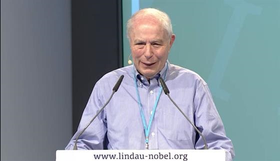
Thank you very much.
Good morning everybody again.
I am very glad to participate in this excellent conference.
It's always a pleasure to interact with young scientists, with students.
I am also learning a lot.
Learning about climate change - thanks a lot, Mario; thanks a lot, Steven.
So it's a great event.
Now what I would like to tell you is about how a discovery is made, using the story of the ubiquitin system as an example.
So I think it is instructive for students and young scientists
to learn how a scientific discovery is made and what can be learned about this.
So I have been interested for a long time in the problem of how proteins are degraded in cells.
And that was a problem that did not interest too many people at that time when I started.
But already at that time, at around 1970, it was known to those who were interested in it
that it is a basically important process.
Abnormal proteins that are made by certain mutations or damages were known to be rapidly eliminated from cells,
which is important for the integrity of cellular function.
But it was known that intracellular protein degradation is not just garbage disposal mechanism to remove abnormal proteins,
because absolutely normal proteins were also known to be selectively degraded at widely different rates.
For example a certain protein was known to be degraded with a half-life time of minutes,
while another took many days or weeks to be degraded.
So it's a highly specific process.
And it was also known that levels of specific proteins, especially the regulatory proteins in animal cells,
can be regulated not only by changes in the rates of synthesis, but also by the changes in the rates of degradation.
That was summarised by Schimke in 1970 when I started to get involved.
So it was known that intracellular...protein degradation of intracellular proteins is a basically important cellular process.
But it was not known how are proteins degraded with such high selectivity and regulation.
So that was the state of the field when I started to work on it.
And I started to work on it when I came as a young postdoctoral fellow - about your age now - to the lab of Gordon Tomkins.
He was working at the University of San Francisco.
He was working at that time on the mechanism by which steroid hormones caused the increased synthesis of enzymes,
specific enzymes in the liver.
So when I got there I saw that he had a big group, 25 postdocs,
and they all worked on different aspects of the synthesis of these enzymes.
I thought this was a bit too congested.
I asked Gordon to give me some other subject.
And he said why don't you work on the degradation of these enzymes, same enzymes.
And that is how I got involved in protein degradation - a subject on which I have been working ever since.
And this slide shows one of the first experiments that I did in the laboratory of Tomkins -
that is the enzyme and that is the degradation.
And I found at that time quite by accident that the degradation of this enzyme is inhibited by potassium fluoride,
an inhibitor of cellular energy production or any other energy production.
So I was very much, even...I should say that this was an accidental finding -
I thought to find the opposite, but that's what I found.
So I was very much impressed by these results, because it said that degradation of intracellular proteins requires energy.
And protein degradation, extracellular protein degradation, such as by a protease as in the gut, actually releases energy.
So I saw that this is a clue:
that there is an energy-dependent system that is responsible for the highly selective degradation of intracellular proteins.
And the question was, what is that system?
So I started to work on this after my postdoctorate fellowship because of the reasons shown in this slide.
As I said protein degradation was known to be important for the control of levels of cellular proteins,
but the biochemical pathways responsible for that were not known.
The energy dependence of protein degradation suggested that there is some kind of a novel unknown biochemical mechanism.
And the very important cause for working on it was that not many were interested, so I did not have to worry about competition.
When I started to work on it after I came back to Israel from my postdoctorate fellowship,
I thought how to go about it and I saw that the best way is to do it by a classical biochemical analysis.
And here is shown what are the stages in the biochemical analysis of the complex system
that gets used for example to find out how protein synthesis or how DNA synthesis works.
First, one has to make a cell-free extract that faithfully reproduces the process in the test tube -
so one has to take apart the cell in order to find what's going on inside.
It's really easy to disrupt the cell, but it's not always easy to disrupt the cell in a way
which will leave you only with an extract that faithfully reproduces the process.
Once one has this, one fractionates the extract because usually it has different enzyme components.
One has to purify each enzyme component to find out how it works, what are the characterisations.
And lastly, the last and important step is to reconstitute the activity of the complete system
by adding back all the isolated and purified components.
And I liken that to trying to find out how a watch, a mechanical watch works.
First you have to open the watch, carefully.
You have to take out the different parts - springs, wheels etc.
So that is what we tried to do.
And our first step was in 1978.
It was published in a small, quite obscure journal, BBRC.
The experiment was done by Aaron Ciechanover who was then my graduate student and whom you heard already.
And what we did was a very simple experiment.
We took lysate from reticulocytes - that was actually reproduced, energy-dependent protein degradation in the test tube.
You can see that the addition of ATP, the cellular energy source, caused a great increase in the degradation of the test protein,
in this case globin.
And then we did a very simple fractionation.
We fractionated this lysate into 2 fractions on an ion exchanger.
Fraction I were all proteins that did not bind to the column, Fraction II where all proteins that bound to the column.
As you can see we lost all protein degradation activity in fraction I.
In fraction II we also lost most of the ATP-stimulated protein degradation.
However, when we combined fraction I with fraction II we got back completely ATP-dependent protein degradation.
So as I said, it was a not too prestigious journal
but I am still fond of this first publication, because that was the first step in the right direction.
It showed us that the system that degrades proteins independence of ATP energy is composed of at least 2 components.
Of course we know now there are many other components.
And as I said, after one fractionate one has to purify and I decided first to purify the active component from fraction II.
And that was a lucky decision because the purification of the active component of fraction II
was the key to the understanding of the mode of the action of the whole system.
So when we purified it, we found that it was a small protein.
It was quite pure.
We called it at that time by the name APF-1 or factor 1 of the system.
And later it was found out to be similar to a previously known protein,
ubiquitin, which was called like that because it is ubiquitous.
They are expressed in all eukaryotic cells, but its function was not known previously.
So the function of ubiquitin is involved somehow in ATP-dependent protein degradation.
And the question was, how?
And the answer came 2 years later - it was quite a big surprise.
We found that ubiquitin is covalently ligated to substrates of the system.
And that is the original experiment here:
We took our purified ubiquitin, labelled it with iodine, incubated it with fraction II in the absence or presence of ATP.
Then electrophoresis in an SDS polyacrylamide gel.
Result:
ATP ubiquitin remains small and ran ahead in the gel.
But following incubation with ATP there was a binding, covalent binding to a huge number of high molecular weight proteins.
And when we saw that we understood that ubiquitin is actually bound to substrate,
or that there is a possibility that it binds to substrates of the system
because crude fraction II contains not only the enzymes but many endogenous substrates of proteolysis.
And to find whether that was indeed the case, we added a known though artificial substrate, lysozyme,
and you can see that there are several additional bands which contain both ubiquitin and lysozyme.
And we analysed them and showed that these are increasing numbers of molecules of ubiquitin bound to one molecule of the substrate.
So based on these findings we proposed in 1980 and we
we proposed that the ligation of ubiquitin or APF-1 here marks proteins for degradation - that is the original scheme.
And we also proposed that there must be some kind of a proteolytic enzyme that after several molecules of ubiquitin are linked to
the protein is degraded to small peptides and then ubiquitin is recycled to be used again.
The main feature...that also gave us the answer to the question, why is energy needed for protein degradation?
It is needed for the tagging mechanism for this - tagging in which the substrate, the protein, is tagged for degradation.
So that was the breakthrough, the original discovery.
And in the following 10 years or so I worked on the intermediary steps in ubiquitin-mediated protein degradation, which gave us...
which showed that the original proposal was actually correct once we added much more important information.
There are 3 enzymes involved in the ligation of ubiquitin to proteins.
Ubiquitin-activating enzyme, a ubiquitin-transferring enzyme or protein E2.
But the main process of the ligation of ubiquitin to specific proteins is done by a class of third enzymes, E3 enzymes,
that recognise certain proteins and then promote the transfer of ubiquitin to form the amide bond with lysine residue of the substrate.
So E3 enzymes are at the heart of the system
because they determine the selectivity and also open the regulation of ubiquitin-mediated protein degradation.
Now more than 600 different E3 enzymes or candidate E3 enzymes are known to be in the human genome.
And we need this large number of ubiquitin protein ligases for the high selectivity of intracellular protein degradation.
And after ubiquitin is ligated and the polyubiquitin chain is formed,
the substrate is degraded by high molecular weight protease complex, 26S proteasome, that was discovered by other people.
And finally 3 ubiquitin is recycled by the action of ubiquitin-C-terminal hydrolases or isopeptidases.
So that was the basic biochemistry of the ubiquitin mediated protein degradation as we found out by about 1990.
And at that time I saw that the time has come to find out what are some important physiological substrates of the system.
And that is how I became interested in the laws of the system, in the cell division cycle.
And that was the first system on which I worked, which is the degradation of cyclin B in the cell cycle.
Cyclin B was discovered by Tim Hunt as a protein that is degraded at the end of each mitosis.
And then it was found that it is the activator, as you all know, is the activator of a kinase, protein kinase Cdk1.
And cyclin B is synthesised in the interface, combines with the kinase, activates it
and then the active protein kinase MPF is formed that promotes mitosis.
And to get out of mitosis, cyclin B has to be degraded.
And that's what happens.
And then the kinase is inactivated and the next cell cycle can take place again.
So I was fascinated with the problem, why is cyclin B stable in the interface
and how it is degraded at the right time at the end of mitosis?
And my approach was again biochemical.
I needed the cell-free system.
And for that I needed another cell-free system.
And that's how I got to this creature, which is a big clam found in the eastern coast of the US.
Usually it is used to make clam chowder.
But it also has some other more scientific uses, mostly to study cell division.
Because a female clam makes hundreds of millions of eggs, and these can be fertilised in the laboratory under control condition;
and then all these millions of eggs begin to divide at a high synchrony.
And what was good for me that one can make extracts at different stages of the cell cycle.
And these extracts, when they are incubated in the test tube,
continue to do faithfully cell-cycle-related events, including cell-cycle-stage-specific cyclin degradation.
So I used these extracts from clam oocytes to find out, how is cyclin degraded at the end of mitosis?
And in 1995 we found or we isolated a large ubiquitin ligase complex that targets cyclins for destruction at the end of mitosis.
And we also got a kind of a rough idea about the mode of its regulation.
And that is taken from this time - we call it the cyclosome, other people called it the anaphase-promoting complex or APC.
And now it is known by these 2 names, anaphase-promoting complex cyclosome or APCC.
And what we propose that it is a large complex.
Now we know it has much more than the number of subunits shown in this original diagram.
But there are a number of subunits, it is not important.
The principle remains that it is inactive in the interface; it gets active at the end of mitosis;
and the initial step to activate it is phosphorylation by its own substrate, by Cdk1 cyclin B.
And once it is phosphorylated there will be some other steps:
It is converted to the active form; and the active form of the APCC, ubiquitin ligase, now ubiquitinates cyclin B,
targets it for degradation, and that inactivates the protein kinase.
And afterwards the ubiquitin ligase is also converted back to the inactive form by dephosphorylation.
So the main idea here is the interrelationship between protein phosphorylation and protein degradation in cell cycle control.
The protein kinase activates the ubiquitin ligase,
and then the active ubiquitin ligase inactivates the protein kinase by the degradation of its activating subunit.
And later it was found that this APCC, which we discovered in clam extracts,
is highly conserved in eukaryotic evolution from yeast to man,
and it has an important, centrally important role in the control of the cell cycle.
So that is very shortly about my own work.
I would like to also briefly mention the work of other people who got into this field.
It turned out that ubiquitin-mediated protein degradation is important in almost every basic cellular function,
such as in the control of cell division - I showed you one example but there are many others -
in the transduction of certain signals, in the regulation of expression of genes
such as those involved in inflammation, immunity, in many stages of embryonic development, in both pro-apoptotic, anti-apoptotic events.
And in all these cases this is done by the timely and regulated degradation of either positive or negative regulators.
So the degradation of regulatory proteins turned out to be very important in cellular regulation.
That looks like energy wasteful -
it really uses up a lot of energy, because you make a protein, you use it only once, then you destroy it.
But by that you make the process essentially irreversible:
so you burn the bridge after you cross it; you cannot go backwards, you can go only forwards.
And of course ubiquitin mediated degradation is also important in protein quality control, as I mentioned,
in getting rid of abnormal proteins.
And since ubiquitin-mediated protein degradation was found to be involved in so many basic processes,
it's not surprising that it is also the cause of many diseases.
And I won't go into the list, I just mention one:
it is the involvement of the ubiquitin system in cancer.
As you know in cancer the cell division gets out of control.
And usually normally cell division is controlled by a balance between the levels of oncoproteins that stimulate cell division,
and of negative regulators, tumour suppressor proteins, that inhibit cell division.
Now in cancer the level of oncoprotein can go up, so you get a very strong stimulation of cell division,
or the level of tumour suppressor protein can go down.
It's like a car with a gas pedal and a brake pedal:
if the gas pedal is put in too much or the brake pedal is not working, the car gets out of control.
Now the important thing from our point of view is that both oncoproteins and tumour suppressor proteins are in a state of...
most of them are in a state of rapid turnover.
So the level of oncoproteins can go down because of decreased degradation.
It is made and synthesised but not degraded.
Or the level of tumour suppresser proteins can go down by increased degradation.
There are many examples of both cases in human cancer.
So the system that we discovered by trying to find out how proteins are degraded turns out to be important in medicine.
And the drug companies are working on it.
There is already one good drug against a certain type of cancer, multiple myeloma, a bone marrow cancer.
It is treated by Velcade or Bortezomib, which is an inhibitor of a proteasome,
and it inhibits the proliferation of myeloma cells, promotes the apoptosis.
It has a very beneficial effect on many cases of multiple myeloma.
It actually revolutionised the treatment of multiple myeloma.
Now you will know that it is a good drug but it's not really a specific drug
because a proteasome inhibitor will inhibit the degradation of all proteins.
It is used in this case in a dose which inhibits only partially the proteasome.
And probably it effects these cancers, especially because myeloma cells make a lot of abnormal proteins.
So the proteasome is overloaded and the further partial inhibition of the proteasome will send them into apoptosis.
But obviously we need more specific agents against other types of abnormalities in cancers or in other diseases.
And for that you need to have more specific aids and target them against specific parts within the system,
such as ubiquitin protein ligases.
So as you see after many years basic research can lead into important applications.
And now I will come to the part that I think I would like to summarise, what can we learn from this story?
What are the lessons from my life in science?
I say 'this far' because I am not yet done.
So I think that my first lesson was that it is very important to have good mentors.
You cannot learn how to do a good science just from reading the literature.
Of course you should read the literature, but you have to talk, you have to interact with mentors.
You have to watch how they do it.
You have to discuss your findings with them, to determine what is the best approach for trying to find your objective.
So to choose very good mentors is very important, and I was very lucky in having very good mentors in my scientific career.
My first mentor was Jacob Mager at the Hebrew University Medical School.
He was a biochemist with encyclopaedic knowledge in all kinds of medical sciences.
He was also a very rigorous experimentalist:
each experiment had to be with all the proper controls, each experiment had to be verified from all possible angles.
So I had a very good solid background of good biochemistry from my work with Jacob Mager.
And that strong biochemistry, solid and rigorous biochemistry, had a great influence on my scientific work ever since.
My second mentor was Gordon Tomkins, whom I mentioned already, at the University of California in San Francisco.
And Gordon was quite different from Mager.
He did not care too much about controls or duplicates, but he had a great imagination.
He was bursting with new ideas; he was a great source of inspiration for many people.
In scienc it is great, it is very important to have imagination, inspiration,
but it is also important not to be carried away with your imagination, but to check rigorously whether your ideas are indeed correct.
So I was very fortunate to combine these 2 mentors:
of imagination, inspiration from Gordon Tomkins, and with the strong, well-controlled biochemistry from Mager.
So my advice to you is: choose good mentors.
If you are not happy with your present mentor, don't hesitate to change him.
My second lesson is to find an important subject that is not yet interesting to others,
otherwise the big guys will get there before you.
Do not go with the mainstream.
So as I told you when I got to the lab of Tomkins, everybody was working on protein synthesis, on synthesis of certain proteins.
It was not only in the lab - at that time protein synthesis was really the main subject,
because that was after the cracking of the genetic code and how proteins are synthesised.
And there was the operon model from Jacob and Monod.
So that was the main interest, how proteins are made.
And not many people were interested in how proteins are degraded.
But I saw that it is important, because proteins are important, and that's a way to regulate protein levels.
So I chose a subject that is not yet interesting to the others,
because if you go with the mainstream you will be the last if you are a young investigator working by yourself.
So try to find a niche that is not yet in the mainstream, but that you believe is biologically important -
they're all important in science if you are a chemist.
Otherwise if it's not important, it's not worth investigating.
Now my third lesson is that accidental observations may be the most important ones.
And when you see an accidental observation you think it's important - don't disregard it, grab your luck!
And as I told you, I did that experiment on the effect of potassium fluoride on the degradation of this enzyme for other reasons.
Actually I thought that it may increase the degradation of the enzyme,
but I found that it stopped and I realised that it was important.
So if you find - of course don't follow any accidental observation -
but if you get an accidental observation which we get almost every time, and you think it's important:
Don't disregard it, grab it, and follow it up.
Number 4 is use whatever experimental approach is needed for to get your objective.
It may not be necessarily the most fashionable or state of the art technology.
So people like to get carried away with the latest technology.
The latest technology is usually powerful.
It's also fashionable.
And there are fashions in science.
And fashion is sometimes good and sometimes not so good.
You should use it only if it is within your objective.
But if it is not useful for your objective, then disregard it and use whatever technique is good for your objective.
So when I started to work on it,
the high-powered techniques of molecular biology that you just heard about, the beginning of them, came into general use.
And as a consequence most biochemists became molecular biologists, and that's fine.
But don't forget your biochemistry too.
So I didn't use it, because you could never find out such a novel mechanism,
like a protein is degraded by being covalently linked to another protein, just by genetic or molecular genetic techniques.
So use whatever technology is most suitable to get your objective.
I also found that science should be...
and when I look back on my life in science I saw how much fun I had actually.
It was a lot of fun, it was a curiosity - really what I mostly did, I tried to satisfy my curiosity.
You can have a lot of excitement and fun.
Now as you will find out we have a lot of chores in science.
We have to collect data in order to write papers.
We have to publish papers in order to get promoted or to get a job.
Or after we get a job to get a grant.
But you shouldn't just collect data to publish papers.
You will survive.
Yes, of course, you have to do that otherwise you won't survive.
If you won't publish, you will perish.
But you shouldn't make your life in science just that:
to publish in order not to perish.
That's not fun that way - then you go to something else.
You should have a lot of excitement.
Until today I am awfully excited whenever there is a new experiment, and I want to know what is the result.
And that is what should drive you, excitement and fun.
Without it you won't make discoveries.
So you should be excited about what you are doing; if you are not excited, leave it.
And my last advice which is not for everybody, but for me it's definitely true:
never to leave bench work and then you shall have to continue to get a lot of excitement and fun.
The people when they get more senior they tend to leave bench work -
I do actually bench work until today, often on a daily basis, and that is my greatest fun.
Also when I do an experiment by my own hands, my connection to the experiment is much more intense.
I am thinking much better trying to find out.
And if it doesn't work, I get very excited and find out what was wrong.
So if it's suitable for you, don't leave bench work and you continue to have a lot of fun and maybe some more discoveries.
But of course you cannot do everything by your own hands, so I want to thank all the people that helped me:
Dvora Ganoth, Hanna Heller, Esther Eytan, Sarah Elias, my wife Judith Hershko who worked in the lab for many years.
I had many graduate students, among them Aaron Ciechanover.
I also collaborated with some very good people, including with Ernie Rose.
And I wanted to show you about these people, one picture how it was at that time.
That was in 1979, in the summer, when I was on a sabbatical at Fox Chase Cancer Center.
And here are some of the people
who were involved in the breakthrough of the discovery of the covalent ligation of ubiquitin to protein.
That was Ernie Rose.
That was Aaron Ciechanover when he was younger.
That was I when I was younger.
So we were all younger then.
Thank you very much for your attention.
Applause.
Vielen Dank und noch einmal einen guten Morgen.
Ich freue mich, dass ich bei dieser ausgezeichneten Veranstaltung dabei sein kann.
Es ist immer eine große Freude, mit jungen Wissenschaftlern, mit Studenten zu tun zu haben.
Auch ich lerne dabei eine Menge.
Ich lerne auch etwas über den Klimawandel.
Vielen Dank dafür, Mario. Vielen Dank, Steven.
Das ist wirklich eine großartige Veranstaltung.
Ich möchte Ihnen etwas darüber erzählen, wie eine Entdeckung erfolgt – am Beispiel der Geschichte des Ubiquitin-Systems.
Für Studenten und Nachwuchswissenschaftler ist es meines Erachtens aufschlussreich zu erfahren,
wie eine wissenschaftliche Entdeckung geschieht und was man daraus lernen kann.
Ich beschäftigte mich bereits seit langem mit der Frage, wie Proteine in Zellen abgebaut werden.
Als ich damals damit begann, interessierte sich kaum jemand für dieses Problem.
Aber bereits damals, so um 1970, wussten Interessierte, dass es sich dabei um einen grundlegend wichtigen Prozess handelt.
Es war bekannt, dass durch bestimmte Mutationen oder Schäden entstehende anormale Proteine schnell aus Zellen entfernt werden.
Das ist wichtig für die Integrität der Zellfunktion.
Man wusste auch, dass der intrazelluläre Proteinabbau nicht nur ein Entsorgungsmechanismus zur Entfernung anormaler Proteine ist,
sondern auch völlig normale Proteine in sehr unterschiedlichen Geschwindigkeiten selektiv abgebaut werden.
So wusste man beispielsweise, dass ein bestimmtes Protein mit einer Halbwertzeit von Minuten abgebaut wird,
während andere dafür Tage oder Wochen benötigen.
Es geht also um einen sehr speziellen Prozess.
Und man wusste auch, dass der Gehalt an Proteinen, insbesondere der regulatorischen Proteine in Tierzellen,
nicht nur durch eine Änderung der Synthesegeschwindigkeiten,
sondern auch durch eine Änderung der Abbaugeschwindigkeiten reguliert werden kann.
Dies hat Schimke 1970 zusammengefasst.
Damals begann ich mich mit dem Thema zu beschäftigen.
Man wusste also, dass der Proteinabbau von intrazellulären Proteinen ein grundlegend bedeutsamer Zellprozess ist.
Nicht bekannt war aber, wie Proteine in einer derartig hohen Selektivität und Regulierung abgebaut werden.
Das war der Stand der Dinge, als ich begann, daran zu arbeiten.
Ich war damals ein junger Postdoc-Mitarbeiter – ungefähr in Ihrem Alter – im Labor von Gordon Tomkins.
Er arbeitete an der University of San Francisco und untersuchte den Mechanismus der Steroidhormone
als Ursache für die vermehrte Synthese von Enzymen, insbesondere Enzymen in der Leber.
Als ich dort anfing, gab es eine große Gruppe, 25 Post-Docs,
die sich mit verschiedenen Aspekten der Synthese dieser Enzyme beschäftigten.
Ich hielt das für etwas überfrachtet.
Deshalb bat ich Gordon, mir ein anderes Thema zuzuweisen.
Und so fragte er mich, ob ich mich nicht mit dem Abbau dieser Enzyme beschäftigen wollte.
Und so kam ich mit dem Thema Proteinabbau in Berührung – ein Thema, an dem ich seitdem arbeite.
Diese Folie zeigt eines der ersten Experimente, das ich im Labor von Tomkins durchführte:
Das ist das Enzym und das ist der Abbau.
Und ich fand damals zufällig heraus, dass der Abbau dieses Enzyms durch Kaliumfluorid,
einen Inhibitor der zellulären Energieproduktion und jeder anderen Energieproduktion, gehemmt wird.
Ich war also sehr … Ich muss sagen, dass das wirklich ein Zufallsergebnis war – ich hatte genau das Gegenteilige erwartet.
Aber das war das Resultat. Ich war sehr beeindruckt von diesen Ergebnissen.
Denn das bedeutete, dass der Abbau intrazellulärer Proteine Energie erfordert.
Und der extrazelluläre Proteinabbau, etwa durch eine Protease im Darm, setzt eigentlich Energie frei.
Für mich war das ein Hinweis auf ein energieabhängiges System,
das für den stark selektiven Abbau von intrazellulären Proteinen verantwortlich ist.
Die Frage war: Um was für ein System handelt es sich?
Nach meinem Postdoc-Stipendiat begann ich aufgrund der in dieser Folie genannten Aspekte mit der Arbeit an diesem Thema.
Man wusste, dass die Proteindegradation für die Steuerung der Menge zellulärer Proteine verantwortlich ist.
Die biochemischen Signalwege, die dafür verantwortlich sind, kannte man jedoch nicht.
Die Energieabhängigkeit des Proteinabbaus ließ vermuten,
dass es so etwas wie einen ungewöhnlichen, unbekannten biochemischen Mechanismus gibt.
Und der entscheidende Grund für meine Arbeit an diesem Thema war die Tatsache,
dass sich kaum jemand dafür interessierte, sodass ich keine Konkurrenz zu fürchten hatte.
Als ich nach dem Postdoc-Stipendiat nach Israel zurückgekehrt war,
begann ich mit der Arbeit und überlegte zunächst die Herangehensweise.
Ich hielt eine klassische biochemische Analyse für den besten Weg.
Hier sehen Sie die Phasen der biochemischen Analyse eines komplexen Systems,
mit der man üblicherweise herauszufinden versucht, wie die Proteinsynthese oder wie die DNA-Synthese funktioniert.
Zunächst muss man ein zellfreier Extrakt herstellen, der den Prozess im Reagenzglas zuverlässig reproduziert.
Man muss die Zelle also auseinandernehmen, um herauszufinden, was im Innern geschieht.
Die Zelle zu zerlegen, ist relativ einfach.
Aber es ist nicht immer einfach, die Zelle so zu zerlegen,
dass man einen Extrakt erhält, der den Prozess zuverlässig reproduziert.
Ist dieser Schritt erledigt, fraktioniert man den Extrakt, weil er normalerweise aus verschiedenen Enzymbestandteilen besteht.
Man muss jeden Enzymbestandteil reinigen, um herauszufinden, wie er funktioniert, welche Merkmale vorhanden sind.
Und schließlich besteht der letzte und wichtigste Schritt darin, die Aktivität des kompletten Systems zu rekonstruieren,
indem man die isolierten und gereinigten Komponenten wieder zusammenfügt.
Ich vergleiche das gerne mit dem Versuch, die Funktionsweise einer mechanischen Uhr zu verstehen.
Zuerst öffnet man die Uhr vorsichtig.
Dann nimmt man die einzelnen Teile, Federn, Rädchen usw., heraus, untersucht sie, setzt sie wieder ein.
Und wenn die Uhr dann läuft, hat man wirklich verstanden, wie das System funktioniert.
Das also haben wir versucht.
Unser erster Schritt erfolgte 1978.
Veröffentlicht wurde das in einer kleinen, ziemlich unbedeutenden Fachzeitschrift, BBRC.
Das Experiment wurde von Aaron Ciechanover durchgeführt, der damals Doktorand bei mir war.
Seinen Vortrag haben Sie ja bereits gehört.
Es handelte sich um ein sehr einfaches Experiment.
Wir nahmen Lysat von Retikulozyten.
Es war ein reproduzierter, energieabhängiger Proteinabbau im Reagenzglas.
Sie sehen, dass die Zugabe von ATP, der zellulären Energiequelle,
den Abbau des Testproteins, in diesem Fall Globin, drastisch verstärkt hat.
Danach haben wir eine sehr einfache Fraktionierung durchgeführt.
Wir haben dieses Lysat in einem Ionenaustauscher in zwei Fraktionen fraktioniert.
Fraktion I bestand aus allen Proteinen, die sich nicht an die Säule gebunden haben,
Fraktion II bestand aus allen Proteinen, die sich an die Säule gebunden haben.
Wie Sie sehen, war die Proteinabbauaktivität in Fraktion I völlig verschwunden.
In Fraktion II war die ATP-stimulierte Proteinabbaureaktion ebenfalls größtenteils verschwunden.
Als wir aber Fraktion I und II zusammenführten, wurde die ATP-abhängige Proteinabbaureaktion vollständig wieder hergestellt.
Es war kein sehr bekanntes Fachjournal.
Aber ich bin immer noch angetan von dieser ersten Veröffentlichung, weil das der erste Schritt in die richtige Richtung war.
Es hat uns gezeigt, dass das System, das Proteine in Abhängigkeit von ATP-Energie abbaut,
aus mindestens zwei Bestandteilen besteht.
Heute wissen wir natürlich, dass es viele weitere Bestandteile gibt.
Wie ich bereits sagte, muss man nach dem Fraktionieren aufreinigen.
Ich beschloss, zunächst den aktiven Bestandteil von Fraktion II zu reinigen.
Und das war eine glückliche Entscheidung,
weil die Aufreinigung des aktiven Bestandteils von Fraktion II der Schlüssel
zum Verständnis des Wirkprinzips des Gesamtsystems war.
Bei der Aufreinigung stellten wir fest, dass es ein kleines Protein war.
Es war ziemlich rein.
Wir bezeichneten das damals als APF-1 oder Faktor 1 des Systems.
Und später stellte sich heraus, dass es einem früher bereits bekannten Protein,
dem Ubiquitin, ähnlich war, das so bezeichnet wurde, weil es ubiquitär ist.
Diese Proteine sind in allen eukaryotischen Zellen exprimiert.
Aber ihre Funktion war vorher unbekannt.
Die Funktion von Ubiquitin ist also irgendwie am ATP-abhängigen Proteinabbau beteiligt.
Die Frage war, wie.
Die Antwort wurde zwei Jahre später gefunden.
Es war eine ziemlich große Überraschung.
Wir fanden nämlich heraus, dass Ubiquitin kovalent an Substrate des Systems gebunden ist.
Hier ist das ursprüngliche Experiment zu sehen:
Wir markierten unser gereinigtes Ubiquitin mit Iodin und inkubierten es – mit oder ohne ATP – mit Fraktion II.
Danach erfolgte eine Elektrophorese in einem SDS-Polyacrylamid-Gel.
Ergebnis: ATP-Ubiquitin blieb gering und setzte sich im Gel an die Spitze.
Aber nach der Inkubation mit ATP bestand eine kovalente Bindung
an eine sehr große Anzahl von Proteinen mit hohem Molekulargewicht.
Und als wir das sahen, begriffen wir, dass Ubiquitin tatsächlich an Substrat gebunden ist
oder eine Bindung an Substrate des Systems möglich ist, weil die rohe Fraktion II nicht nur die Enzyme,
sondern viele endogene Proteolysesubstrate enthält.
Und um herauszufinden, ob das tatsächlich stimmt, gaben wir ein bekanntes, aber künstliches Substrat, Lysozym, hinzu.
Und Sie sehen hier mehrere zusätzliche Bänder, die sowohl Ubiquitin als auch Lysozym enthalten.
Wir haben das analysiert und nachgewiesen, dass es sich hierbei um zunehmende Anzahlen von Molekülen des Ubiquitins handelt,
die an ein Molekül des Substrats gebunden sind.
Auf der Grundlage dieser Ergebnisse entwickelten wir
und meinem Kollegen vom Fox Chase Cancer Center, Irwin Rose – 1980 das Konzept,
dass die Bindung von Ubiquitin oder APF-1 Proteine für den Abbau markiert.
Das ist das ursprüngliche Schema.
Wir gingen auch davon aus, dass es eine Art von proteolytischem Enzym geben muss,
das in kleine Peptide abgebaut wird, nachdem sich mehrere Ubiquitin-Moleküle an das Protein gebunden haben.
Danach wird das Ubiquitin wieder verwendet.
Das Hauptmerkmal beantwortete auch unsere Frage, warum für den Proteinabbau Energie benötigt wird.
Sie wird für den darin enthaltenen Markierungsmechanismus benötigt,
bei dem das Substrat, das Protein, das abgebaut werden soll, markiert wird.
Das war der Durchbruch, die ursprüngliche Entdeckung.
In den nachfolgenden zehn Jahren habe ich die Zwischenschritte beim Ubiquitin-vermittelten Proteinabbau untersucht.
Dabei zeigte sich, dass die ursprüngliche Vermutung tatsächlich stimmte.
Wir konnten dieses Ergebnis durch weitere wichtige Informationen ergänzen.
An der Bindung von Ubiquitin an Proteinen sind drei Enzyme beteiligt,
nämlich ein Ubiquitin-aktivierendes Enzym, ein Ubiquitin-übertragendes Enzym oder Protein E2.
Aber der hauptsächliche Prozess der Bindung von Ubiquitin an spezielle Proteine
erfolgt durch eine dritte Enzym-Kategorie, E3-Enzyme, die bestimmte Proteine erkennen
und dann die Übertragung von Ubiquitin fördern, um die Amid-Bindung an den Lysinrest des Substrats zu bilden.
Im Zentrum des Systems stehen also E3-Enzyme.
Sie entscheiden über die Selektivität und leiten die Regulierung des Ubiquitin-vermittelten Proteinabbaus ein.
Inzwischen sind über 600 verschiedene E3-Enzyme oder potenzielle E3-Enzyme im Humangenom bekannt.
Wir benötigen diese riesige Anzahl von Ubiquitin-Protein-Ligasen für die hohe Selektivität des intrazellulären Proteinabbaus.
Nachdem das Ubiquitin gebunden ist und die Polyubiquitinkette gebildet wurde,
wird das Substrat durch einen Proteasekomplex mit hohem Molekulargewicht, 26S-Proteasom, abgebaut, den andere entdeckt haben.
Und schließlich wird 3-Ubiquitin durch die Aktion von C-terminierten Ubiquitin-Hydrolasen oder Isopeptidasen recycelt.
Das war die grundlegende Biochemie des Ubiquitin-vermittelten Proteinabbaus, wie wir sie bis rund 1990 untersucht hatten.
Zum damaligen Zeitpunkt dachte ich, dass es an der Zeit wäre, einige bedeutende physiologische Substrate des Systems aufzuklären.
Deshalb interessierte ich mich für die Gesetzmäßigkeiten des Systems, den Zellteilungszyklus.
Das war das erste System, über das ich arbeitete.
Es ging um den Abbau von Cyclin B im Zellzyklus.
Cyclin B wurde von Tim Hunt als ein Protein entdeckt, das zum Ende jeder Mitose abgebaut wird.
Und dann fand man heraus, dass es der Aktivator einer Kinase, nämlich von Proteinkinase Cdk 1, ist.
Cyclin B wird an der Schnittstelle synthetisiert, schließt sich mit der Kinase zusammen,
aktiviert sie und dann bildet sich die aktive Proteinkinase MPF, die die Mitose fördert.
Und um die Mitose zu beenden, muss Cyclin B abgebaut werden.
Und genau das geschieht.
Die Kinase wird inaktiviert und der nächste Zellzyklus kann erfolgen.
Mich faszinierte die Frage, warum Cyclin B an der Schnittstelle stabil ist
und wie es am Ende der Mitose zum richtigen Zeitpunkt abgebaut wird.
Und auch hier verfolgte ich einen biochemischen Ansatz.
Ich benötigte das zellfreie System.
Und dafür brauchte ich ein weiteres zellfreies System.
Dabei stieß ich auf diese Kreatur, eine Riesenmuschel, die an der Ostküste der USA vorkommt.
Normalerweise verwendet man sie für eine Muschelsuppe.
Aber sie lässt sich auch zu wissenschaftlichen Zwecken nutzen, hauptsächlich zur Untersuchung der Zellteilung.
Denn eine weibliche Muschel erzeugt hunderte Millionen Eier,
die im Labor unter kontrollierten Bedingungen befruchtet werden können.
Und dann teilen sich diese Millionen von Eiern mit einer hohen Synchronizität.
Der Vorteil für mich war, dass sich Extrakte aus unterschiedlichen Phasen des Zellzyklus gewinnen lassen.
Und bei Inkubation im Reagenzglas spulen diese Extrakte weiterhin für den Zellzyklus relevante Schritte ab,
auch den für die Zellzyklusphase spezifischen Cyclin-Abbauprozess.
Ich nahm also diese Extrakte aus den Muscheleizellen, um herauszufinden,
wie Cyclin am Ende der Mitose abgebaut wird.
Und 1995 fanden wir bzw. isolierten wir einen riesigen Ubiquitin-Ligasekomplex,
der auf die Zerstörung von Cyclinen am Ende der Mitose abzielt.
Und wir hatten auch eine ungefähre Vorstellung darüber, wie dieser Vorgang reguliert wird.
Diese Aufnahme stammt aus dieser Zeit.
Wir nennen das das Cyclosom.
Andere nennen das den Anaphase Promoting Complex oder APC.
Und heute kennt man das unter diesen Bezeichnungen: Anaphase Promoting Complex/Cyclosom oder APCC.
Und wir denken, dass es ein riesiger Komplex ist.
Inzwischen wissen wir, dass es mehr Untereinheiten gibt, als in dieser ursprünglichen Darstellung zu sehen ist.
Es gibt also zahlreiche Untereinheiten.
Das ist aber nicht so wichtig.
Der Grundsatz bleibt derselbe, dass nämlich der Komplex an der Schnittstelle inaktiv ist.
Am Ende der Mitose wird er aktiviert.
Der erste Schritt der Aktivierung ist die Phosphorylierung durch sein eigenes Substrat, Cdk 1 Cyclin B.
Nach der Phosphorylierung erfolgen weitere Schritte: Der Komplex wird in die aktive Form umgewandelt.
Die aktive Form von APCC, Ubiquitin-Ligase, ubiquitiniert jetzt Cyclin B und ist auf den Abbau von Cyclin B gerichtet.
Dadurch wird die Proteinkinase inaktiviert.
Und anschließend wird die Ubiquitin-Ligase durch Dephosphorylierung ebenfalls in die inaktive Form zurückverwandelt.
Das Grundkonzept dabei ist die Wechselwirkung zwischen Proteinphosphorylierung und Proteinabbau in der Kontrolle des Zellzyklus.
Die Proteinkinase aktiviert die Ubiquitin-Ligase
und die aktive Ubiquitin-Ligase inaktiviert die Proteinkinase durch Abbau ihrer aktivierenden Untereinheit.
Später wurde festgestellt, dass dieser APCC, den wir in Muschelextrakten entdeckt hatten,
in der eukaryotischen Entwicklung von der Hefe zum Menschen konserviert wurde
und eine zentrale Rolle in der Zellzykluskontrolle spielt.
Das war eine sehr kurz gefasste Übersicht über meine eigenen Arbeiten.
Ich möchte auch kurz die Arbeiten anderer Menschen erwähnen, die auf diesem Gebiet aktiv waren.
Es hat sich herausgestellt,
dass der Ubiquitin-vermittelte Proteinabbau für fast jede grundlegende zelluläre Funktion Bedeutung hat:
etwa in der Kontrolle der Zellteilung – ich habe Ihnen ein Beispiel gezeigt
beispielsweise von Genen, die an Entzündungsprozessen und Immunität beteiligt sind,
in vielen Phasen der Embryonenentwicklung sowie in pro-apoptotischen als auch anti-apoptotischen Ereignissen.
Diese Vorgänge erfolgen alle durch den rechtzeitigen und regulierten Abbau entweder positiver oder negativer Regulatoren.
Der Abbau regulatorischer Proteine hat sich als sehr wichtig in der Zellenregulierung erwiesen.
Das sieht nach purer Energieverschwendung aus.
Dafür wird tatsächlich eine Menge Energie verbraucht.
Denn es wird ein Protein hergestellt, das nur einmal verwendet und dann zerstört wird.
Aber dadurch wird der Prozess im Wesentlichen unumkehrbar: Man reißt die Brücke ein, nachdem man sie überquert hat.
Eine Umkehr ist nicht möglich, es geht nur vorwärts.
Der Ubiquitin-vermittelte Abbau hat, wie gesagt,
natürlich auch Bedeutung für die Steuerung der Proteinqualität, für die Beseitigung anormaler Proteine.
Und da der Ubiquitin-vermittelte Proteinabbau nachweislich an so vielen grundlegenden Prozessen beteiligt ist,
überrascht es nicht, dass er auch die Ursache vieler Krankheiten ist.
Und ich möchte sie hier nicht alle aufführen, aber einen Aspekt nennen:
die Beteiligung des Ubiquitin-Systems an Krebserkrankungen.
Bekanntlich gerät die Zellteilung bei Krebs außer Kontrolle.
Normalerweise wird die Zellteilung gesteuert durch ein ausgeglichenes Verhältnis zwischen der Menge von Onkoproteinen,
die die Zellteilung stimulieren, und von negativen Regulatoren, Tumorsuppressor-Proteinen,
die die Zellteilung verhindern.
Bei Krebs kann die Menge an Onkoproteinen ansteigen, sodass die Zellteilung sehr stark stimuliert wird,
oder die Menge der Tumorsuppressor-Proteine kann abnehmen.
Das ist wie mit dem Gas- und Bremspedal beim Auto:
Wenn das Gaspedal zu stark durchgedrückt wird oder das Bremspedal nicht funktioniert, gerät das Fahrzeug außer Kontrolle.
Aus unserer Sicht ist dabei entscheidend,
dass sowohl die meisten Onkoproteine als auch die meisten Tumorsuppressor-Proteine schnell umgesetzt werden.
Die Menge an Onkoproteinen kann also aufgrund eines verminderten Abbaus abnehmen.
Die Proteine werden produziert und synthetisiert, aber nicht abgebaut.
Oder die Menge an Tumorsuppressor-Proteinen kann aufgrund eines zunehmenden Abbaus abnehmen.
Bei menschlichen Krebserkrankungen gibt es viele Beispiele für beide Varianten.
Das von uns bei dem Versuch, den Abbau von Proteinen zu verstehen,
entdeckte System spielt eine entscheidende Rolle in der Medizin.
Und die Pharmazieunternehmen arbeiten daran.
Es gibt bereits ein gutes Arzneimittel gegen einen bestimmten Krebs, das multiple Myelom, ein Knochenmarkkrebs.
Er wird mit Velcade oder Bortezomib behandelt.
Das ist ein Inhibitor eines Proteasoms.
Er verhindert die Verbreitung von Myelomzellen und fördert die Apoptose.
Er hat sehr positive Auswirkungen auf multiple Myelome und die Behandlung des multiplen Myeloms wirklich revolutioniert.
Nun weiß man zwar, dass das ein gutes Arzneimittel ist,
aber kein gezielt einsetzbares Arzneimittel, da ein Proteasom-Inhibitor den Abbau aller Proteine verhindert.
Er wird in diesem Fall in einer Dosis verabreicht, die das Proteasom nur teilweise hemmt.
Und wahrscheinlich ist es bei diesen Krebsarten besonders wirksam, weil Myelomzellen eine Menge anormaler Proteine erzeugen.
Das Proteasom ist also überlastet und die partielle Hemmung des Proteasoms löst ihre Apoptose aus.
Aber selbstverständlich brauchen wir weitere spezielle Wirkmittel
gegen andere Arten von Anomalitäten bei Krebs oder anderen Erkrankungen.
Und dazu werden stärker spezialisierte Mittel benötigt,
die man gezielt gegen spezielle Teile des Systems einsetzen kann, wie Ubiquitin-Protein-Ligasen.
Viele Jahre Grundlagenforschung können also, wie Sie sehen, zu bedeutenden Anwendungen führen.
Und jetzt möchte ich zusammenfassen, was man aus dieser Geschichte lernen kann.
Welche Lektionen habe ich in meinem wissenschaftlichen Leben gelernt?
Ich sage „bisher gelernt“, weil ich ja noch nicht am Ende bin.
Meine erste Lektion ist die, dass gute Mentoren enorm wichtig sind.
Man lernt nicht, gute Wissenschaft zu betreiben, indem man einfach nur Fachliteratur liest.
Natürlich muss man die Literatur lesen.
Aber man muss auch mit Mentoren sprechen, sich mit ihnen austauschen.
Man muss beobachten, wie sie arbeiten.
Man muss eigene Ergebnisse mit ihnen diskutieren, um herauszufinden, wie man sein Ziel am besten definiert.
Wichtig ist also die Wahl guter Mentoren.
Ich hatte in meiner wissenschaftlichen Karriere das Glück, sehr gute Mentoren zu haben.
Mein erster Lehrmeister war Jacob Mager an der Medizinischen Hochschule der Hebräischen Universität in Jerusalem.
Er war Biochemiker und verfügte in allen Bereichen der medizinischen Forschung über enzyklopädisches Wissen.
Zugleich war er rigoroser Experimentalist: Jedes Experiment musste mit sachgemäßen Kontrollen durchgeführt werden.
Jedes Experiment musste aus allen möglichen Blickwinkeln überprüft werden.
Aus meiner Arbeit mit Jacob Mager stammt der sehr gute und solide Hintergrund guter Biochemie.
Und diese solide und rigorose Biochemie hat meine wissenschaftliche Arbeit seitdem stark geprägt.
Mein zweiter Mentor war der bereits erwähnte Gordon Tomkins an der University of California in San Francisco.
Gordon war ganz anders als Mager.
Er legte nicht so viel Wert auf Kontrollen oder Duplikate.
Er verfügte aber über eine große Imaginationskraft.
Er sprühte nur so vor Ideen und hat viele Menschen enorm inspiriert.
In der Wissenschaft sind Imaginationskraft und Inspiration sehr von Vorteil.
Wichtig ist aber auch, sich nicht von der eigenen Vorstellungskraft wegtragen zu lassen,
sondern rigoros zu überprüfen, ob die eigenen Ideen tatsächlich stimmig sind.
Deshalb war es für mich ein besonderes Glück, die Einflüsse dieser beiden Mentoren kombinieren zu können:
die Imagination und die Inspiration von Gordon Tomkins und die solide, gut überprüfte Biochemie von Mager.
Ich empfehle Ihnen also, sich gute Mentoren zu wählen.
Wenn Sie mit Ihrem aktuellen Mentor unzufrieden sind, sollten Sie nicht zögern, zu wechseln.
Die zweite Lektion, die ich gelernt habe, ist die, ein wichtiges Thema zu finden,
das für andere nicht interessant genug ist.
Denn sonst sind die anderen vor mir am Ziel.
Gehen Sie nicht mit dem Mainstream.
Zu dem Zeitpunkt, als ich in das Labor von Tomkins wechselte,
arbeiteten alle zur Proteinsynthese, zur Synthese bestimmter Proteine.
Nicht nur in diesem Labor.
Damals war die Proteinsynthese wirklich das zentrale Thema,
weil der genetische Code gerade geknackt worden war und man Proteine synthetisieren konnte.
Und es gab das Operon-Modell von Jacob und Monod.
Es war Hauptthema, wie Proteine hergestellt werden.
Kaum jemand interessierte sich dafür, wie Proteine abgebaut werden.
Aber ich erkannte die Bedeutung, weil Proteine wichtig sind und der Abbau eine Möglichkeit ist, den Proteingehalt zu regulieren.
Ich wählte also ein für andere uninteressantes Thema.
Wenn man als junger, allein arbeitender Nachwuchsforscher mit dem Mainstream geht, ist man das Schlusslicht.
Versuchen Sie also eine Nische zu finden, die noch nicht im Mainstream angekommen ist,
aber die Sie für biologisch relevant halten.
Für einen Chemiker sind alle wissenschaftlichen Themen von Bedeutung.
Wenn es aber nicht relevant ist, lohnt sich auch die Beschäftigung damit nicht.
Meine dritte Lektion ist die, dass zufällige Beobachtungen möglicherweise die wichtigsten sind.
Wenn Sie eine zufallsbedingte Beobachtung machen, sollten Sie sie für wichtig halten.
Missachten Sie sie nicht.
Versuchen Sie Ihr Glück!
Wie ich bereits sagte, habe ich das Experiment über die Auswirkung von Kaliumfluorid auf den Abbau dieses Enzyms
aus ganz anderen Gründen durchgeführt.
Ich hatte erwartet, dass es den Abbau des Enzyms verstärkt,
fand aber heraus, dass es den Abbau hemmt, und stellte fest, dass das von Bedeutung ist.
Sollten Sie also einmal eine zufallsbedingte Beobachtung machen – natürlich folgt man nicht jeder derartigen Beobachtung –
und diese für bedeutsam halten: Missachten Sie sie nicht, sondern greifen Sie sie auf und verfolgen Sie sie weiter.
Lektion Nummer 4 lautet: Nutzen Sie jeden experimentellen Ansatz, der für das Erreichen Ihres Ziels notwendig ist.
Es muss nicht immer die modernste oder neueste Technologie sein.
In der neuesten Technologie kann man sich leicht verlieren.
Die neueste Technologie ist oft sehr einflussreich.
Sie ist auch modern.
Es gibt auch in der Wissenschaft Moden.
Und jede Mode hat etwas Gutes und etwas weniger Gutes.
Sie sollten sie nur einsetzen, wenn sie zu Ihrem Ziel passt.
Wenn Sie aber für Ihr Ziel nicht sinnvoll ist, missachten Sie sie und nutzen Sie eine Technik, die gut für Ihr Ziel ist.
Als ich an dem Thema zu arbeiten begann,
fanden gerade die leistungsstarken Techniken der Molekularbiologie Einzug, von denen Sie gerade gehört haben.
Und deshalb wurden viele Biochemiker Molekularbiologen und das ist gut so.
Aber vergessen Sie die Biochemie nicht.
Ich habe die Molekularbiologie nicht eingesetzt,
weil man einen so ungewöhnlichen Mechanismus wie den Proteinabbau
durch kovalente Bindung an ein anderes Protein niemals ausschließlich
durch genetische oder molekulargenetische Techniken entdecken könnte.
Setzen Sie also die Technologie ein, die für Ihr Ziel am besten geeignet ist.
Ich war auch immer der Meinung, dass Wissenschaft … Wenn ich auf mein Leben in der Wissenschaft zurückblicke,
stelle ich fest, wie viel Spaß mir das tatsächlich gemacht hat.
Es war Freude, es war Neugier – tatsächlich habe ich überwiegend dafür gesorgt, meine Neugier zu befriedigen.
Und dabei kann man eine Menge Begeisterung und Spaß entwickeln.
Wie Sie erleben werden, gibt es auch in der Wissenschaft jede Menge lästige Pflichten.
Wir müssen Daten sammeln, um Artikel zu schreiben.
Wir müssen Artikel veröffentlichen, um unterstützt zu werden oder einen Job zu erhalten oder,
wenn wir einen Job haben, um finanzielle Fördermittel zu erhalten.
Aber man sollte nicht einfach Daten sammeln, um veröffentlichen zu können.
Dann überlebt man. Ja, natürlich müssen Sie das machen, weil Sie sonst nicht überleben könnten.
Wer nicht veröffentlicht, geht unter.
Aber Sie sollten Ihr Leben in der Wissenschaft nicht so gestalten, dass Sie veröffentlichen, um nicht unterzugehen.
Das macht keinen Spaß. Dann führt Sie Ihr Weg irgendwo anders hin.
Sie sollten eine Menge Begeisterung mitbringen.
Bis heute bin ich furchtbar aufgeregt, wann immer es ein neues Experiment gibt, und gespannt auf das Ergebnis.
Und genau das sollte Sie antreiben: Begeisterung und Freude.
Ohne das werden Sie keine Entdeckungen machen.
Sie sollten begeistert von dem sein, was Sie tun.
Wenn Sie nicht begeistert sind, lassen Sie es sein.
Und mein letzter Rat ist sicherlich nicht für jeden gedacht, aber definitiv für mich.
Hören Sie nie mit der Laborarbeit auf.
Dann werden Sie einfach eine Menge Begeisterung und Freude erleben.
Menschen tendieren mit zunehmendem Alter dazu, die Laborarbeit aufzugeben.
Ich mache bis heute Laborarbeit, oft jeden Tag.
Das ist meine größte Freude.
Wenn ich mit meinen eigenen Händen ein Experiment durchführe, ist meine Verbindung zum Experiment wesentlich intensiver.
Ich kann viel besser nachdenken, wenn ich die Lösung ausprobiere.
Und wenn das nicht funktioniert, bin ich ziemlich aufgeregt und möchte unbedingt herausfinden, was falsch war.
Wenn es also für Sie stimmig ist,
sollten Sie die Laborarbeit nie aufgeben und Sie werden eine Menge Freude und möglicherweise einige Entdeckungen mehr erleben.
Aber natürlich können Sie auch nicht alles selbst machen, weshalb ich auch all den Menschen danken möchte,
die mich unterstützt haben: Dvora Ganoth, Hanna Heller, Esther Eytan, Sarah Elias, meiner Frau Judith Hershko,
die viele Jahre im Labor gearbeitet haben.
Ich hatte viele Doktoranden, unter ihnen Aaron Ciechanover.
Und ich habe auch mit einigen sehr guten Leuten zusammengearbeitet, zum Beispiel mit Ernie Rose.
Ich möchte Ihnen ein Bild aus der Arbeit mit diesen Menschen aus der damaligen Zeit zeigen.
Es war im Sommer 1979, als ich einen Studienurlaub im Fox Chase Cancer Center verbrachte.
Und hier sind einige der Menschen,
die an der bahnbrechenden Entdeckung der kovalenten Bindung von Ubiquitin an Protein beteiligt waren.
Das war Ernie Rose.
Das war Aaron Ciechanover, als er jünger war.
Das war ich, als ich jünger war.
Wir alle waren damals etwas jünger.
Vielen Dank für Ihre Aufmerksamkeit
Applaus.