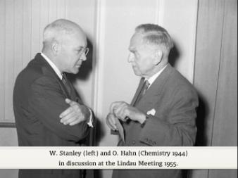
It is a pleasure to see so many of you here this morning after the very pleasant evening that we all enjoyed last night.
I want to take this opportunity to express my thanks to the group at Lindau
for inviting me and Mrs. Stanley to attend this meeting.
Fortunately it fitted in with a trip through Europe in company with our three daughters.
They too are enjoying this bit of hospitality.
I can only say that it reminds me very much of an earlier pleasant experience in Stockholm which Mrs. Stanley and I enjoyed.
Lindau it seems to me is for the time being just a little bit of Sweden.
Now, to the topic of viruses.
I have considerable difficulty in determining the nature of the talk this morning
because we range here in the audience from experts in a variety of specialities to, as I understand, beginning students.
This makes it somewhat difficult.
I shall attempt therefore to give a sort of cross section of the virus work
carried out in our laboratory at the University of California, in Berkeley.
First I shall attempt just a little bit of background material of philosophy if you please.
Then a little bit of chemistry, then some current work on poliomyelitis virus and vaccine.
And hope that you will find something of interest in one or another of these divisions.
Now, viruses represent a comparatively new field since they were discovered just at the turn of the century.
The first virus to be discovered was that of tobacco mosaic, a plant virus.
The next was an animal virus, that of the foot and mouth disease of cattle.
The third was the virus of yellow fever discovered by Reed and co-workers in 1901.
For about 30 years the work on viruses stemmed largely on medical aspects, upon the disease producing characteristics of viruses.
Very little was known about the true characters of these infectious disease producing agents.
Now, perhaps a definition of a virus at the outset.
They are small, extremely small, self duplicating mechanisms
which multiply or reproduce only within the living cells of certain specific hosts.
During their multiplication or their growth or their reproduction in these living cells they occasionally change or mutate.
And when they do this, they produce a new kind of a disease.
Hence the viruses provide an unusually fruitful pathway to the area that Professor Muller will speak about later.
Because of the ability to reproduce or to grow, because of the ability of viruses to mutate or to change,
they have for years been regarded as examples of life.
Now, in order to understand what one speaks about when one talks about a living entity,
it is necessary to indulge in a bit of philosophy with respect to the nature of life.
You have no difficulty in seeing the persons surrounding you as examples of living entities.
And yet the metal of the microphone you have as an admitted example of a non living structure.
Yet somewhere in between there is a borderline, a line of the unknown so to speak,
a boundary between living things and non-living things.
It is of some interest that an early philosopher, Aristotle, thought about this boundary line
and about 2,000 years ago suggested that the boundary line between living things and non-living things
was doubtful and perhaps non-existent.
So now you may ask where do the viruses fit in to this size of structures which exists in the world?
And I have prepared a slide which I hope will give you a better idea of this borderline area.
By 1930 chemists had worked up to the large molecules, some of which you heard Professor Staudinger discuss yesterday.
The large macromolecules of the proteins which on this scale will come to somewhere in this range.
In other words, chemists working with larger and larger and larger molecules worked up to protein molecules,
for example those of the hemocyanin molecule.
And beyond this there was the gap of the unknown.
The biologists on the other hand working downwards from larger and larger animals
had come down to the second line from the top, that is bacillus prodigiosus, for example.
The bacteria, which on this scale are in the neighbourhood of 450 or so millimicrons,
and between the bacteria or the smallest of the living organisms of the biologist
and the largest of the chemical molecules of the chemists, there was this unknown area.
This is the area I think that Aristotle was speaking,
that the boundary line in between the two was doubtful and perhaps non-existent.
Prior to the discovery of the viruses, this area was an unknown area.
But, as you can see, many, many, many entities have been filled in here,
so that if you look at the size of the structure, going from the smallest here to the largest there,
there is almost a continuum with respect to structures.
So that in truth there is no boundary line now between the accepted living organisms of the biologist,
such as bacillus prodigiosus and the molecules of the chemists at this level.
And the viruses, these infectious disease producing agents have been the structures
which serve to fill in at long last this gap which has existed for years and years.
Well, now I indicated that from the time of the discovery of viruses, around the turn of the century,
up until about 1935, the basic nature of these structures in this area was unknown.
It was not known whether they were still smaller ordinary living organisms like bacillus prodigiosus.
Some new kind of a chemical molecule or simply a specialised grouping attached to a chemical molecule.
And it became very important to determine the exact nature of one of the viruses.
And for this, the virus in the middle here, tobacco mosaic virus was selected for chemical study.
And to make a long story quite short, this virus was isolated in the form of a crystallisable nucleoprotein.
And after a long series of tests designed to prove whether or not the biological activity
was a part and parcel of the protein, the nucleoprotein molecule,
it was concluded that beyond a reasonable doubt the virus activity was a specific property of the nucleoprotein.
Then came of course the business of finding out whether tobacco mosaic virus was a representative virus
or whether it was something unusual.
So studies on a variety of viruses were made.
And for example another virus was obtained in the form of the beautiful dodecahedral crystals that you see here.
This is another plant virus, that of the tomato bushy stunt virus.
It can be isolated in the form of the crystals that you see here.
These crystals are composed of very tiny macromolecules about 30 or so millimicrons in diameter.
And then, to give you a cross section now of the variety of structures that have been isolated
in the form of purified viruses, I’d like to show this slide,
which in effect covers almost the entire range of sizes of the viruses that I showed you on the first chart of sizes.
Starting at the top upper left, we have the vaccinia, elementary bodies of vaccinia,
the vaccine which is used to protect you against smallpox, you will probably all have had the little scratch on your arm.
Probably didn’t realise that they were rubbing into your arm the large redshaped objects that you see on the upper right hand.
At the upper right we have an example of a smaller virus, that of influenza virus.
An extremely interesting virus, one which could serve as the subject of an entire lecture,
because this virus in 1918 caused the deaths of more people than had died on the battle fields of two great world wars.
In our own country of the United States we lost over 400,000 persons within a 4 month period
and activities of that virus are on the upper right hand side.
And in 1918 influenza was not even recognised as a virus disease, for it was not so discovered until 1931,
first by the English workers and a similar virus in swine by Doctor Shope in our laboratory.
Here is an example of a group of viruses known as bacteria viruses, the viruses which attack bacteria.
Here is a T2 bacterial virus with an unusual structure, such as you see, with the head and the sperm shape tail.
On the right hand side here is an extremely interesting virus because this is a cancer producing virus.
A virus which produces cancer in rabbits in the United States.
It is of course of extreme interest
because this provides one of the avenues that approached to the solution of the cancer problem.
This is another example of a bacteria virus or a bacterial virus.
This one has the very short stubby tail, this tail organ is very important
because it is believed that this provides a mechanism by means of which the infective process is carried out.
And for some time it was not recognised that this particular bacteria virus had a tail.
Here is an example of the little macromolecules which go to make up the tomato bushy stunt virus,
which gives the beautiful crystals that you saw just a moment ago.
Here is a familiar tobacco mosaic virus about which I shall say just a bit more in a moment.
And here are some molecules, macromolecules perhaps is a better word, of a virus which affects orchids.
You ladies perhaps have not realised that the flowers that you wear on your dresses in the evening,
the orchids are subject also to virus infection.
And when you isolate the virus which causes a disease of orchids, you obtain the material on the right.
Now, if you go from the small spherical virus through the range of sizes that you see here,
you cover almost the entire range from about 20 millimicrons up to 300 millimicrons.
So that here you have a birds eye view of real structures
which have been found to exist in this borderline which existed between the living and the non-living.
Now, for just a little bit of chemistry, needless to say my background and my training is in chemistry.
Having isolated some of these materials such as tobacco mosaic viruses,
it was only natural to subject purified preparations of this virus to the normal procedures of characterisation,
analysis and so forth.
And the next slide will give you an example of the building blocks which go to make up the tobacco mosaic virus.
Now, for those of you who are not chemists, don’t worry too much, I’ll point out in gross terms the significance of this slide.
But first this row or columns give the amounts of the various amino acids which go to make up the tobacco mosaic virus.
You can see them here.
These amounts are very characteristic,
whether you isolate the tobacco mosaic virus in Sweden, in the United States, in Australia,
whether you isolate the virus from Turkish tobacco plants, from tomato plants, from spinach plants.
In other words, there is a characteristic composition which exists throughout the world
and regardless of the host in which you grow this material.
Now, I indicated at the beginning of this talk that one of the characteristics of viruses is that they can mutate.
Needless to say as chemists we wondered what would happen when a virus mutates and causes a slightly different kind of a disease.
We therefore isolated it and purified a variety of strains of tobacco mosaic virus.
And the results of these analysis are also on this slide.
We have the masked strain of the virus, the JD141 strain of the virus,
the green aucuba, the yellow aucuba, the ribgrass virus and two cucumber viruses.
These for a time have been regarded as strains of tobacco mosaic virus.
Now, whenever differences in composition exist, differences in composition from the tobacco mosaic virus,
the figure has been imposed in a little block.
So as I have indicated, those of you who are not chemists,
you need only to determine the number of little figures enclosed in a heavy line
to get some idea of the nature and extent of the changes which exist.
For example, there are only two changes in the JD141 strain, here and here.
On the other hand, in the case of the Holmes ribgrass virus, there are many, many changes in the nature of the amino acids.
And as you can see, there is an example here of the introduction of a new amino acid into the strain.
In other words, the amino acid or the building block, if you please, histidine does not exist in the tobacco mosaic virus.
Yet when this strain mutated and formed the Holmes ribgrass strain,
it was accompanied by the introduction of histidine, it was accompanied by the introduction of a new amino acid.
This data provided therefore the first information concerning the nature of mutation in the field of viruses.
And I hope this may also serve as an indication of the nature
of the changes which take place in the mutation of genes of higher organisms.
And if so, this provides an experimental approach to the nature of the changes
that Professor Muller has been interested in these many years.
Now, we have recently been interested in the detailed structure of tobacco mosaic virus.
You can make a variety of kinds of studies on this virus
and one of the useful approaches from a standpoint of technique
is the newly developed spray drop technique of Professor Williams in our laboratory.
In which a solution of virus is mixed in known proportions with a polystyrene latex solution.
The polystyrene latex particles are shown here and the tobacco mosaic virus particles are here.
And you see a small segment at the right which has been enlarged, so that you can see the particles somewhere on the plain.
By mixing the two, you get a relationship between the number of particles of polystyrene latex
which you know by virtue of having prepared the solution and the number of particles of tobacco mosaic virus.
You get a micro drop and hence a representative sample of this mixture by means of a spraying technique from an atomiser.
And the spray drop is showing an outline here, shows quite clearly.
Hence, this micro drop gives you a representative sample and enables you to get a relationship
between the number of particles of tobacco mosaic virus and the number of particles of the polystyrene latex.
And thus you can carry out much work on the relationship of biological activity to the little rods which go to make up the virus.
Now, our recent work on the structure, the detailed structure of these rods, has given us some rather unusual results.
This work was stimulated primarily by the results obtained in our laboratory and in other laboratories
on the bacteriophage particles.
And work, stemming also from the workers here in Germany on a protein component
which can be obtained by degradation of tobacco mosaic virus.
The next slide shows in outline some of the electron micrographs which can be obtained from such mixtures.
At the upper left in high magnification is the intact tobacco mosaic virus particle.
And if you break this particle, for example by means of high sound treatment,
you can simply slice it across as you would slice a piece of sausage.
If you do that, you get the two particles, or you get particles such as those that you see in the upper right hand corner.
These have been found to be hexagons so it would appear that the cross section of this rod is that of a hexagon.
Within the past few months it has been possible to devise a technique
by means of which an individual macromolecule can be partially denatured.
If you subject a solution of tobacco mosaic virus to mild temperature treatment, to mild heat, the one end will tend to ball up.
And when this is then treated with a detergent such as sodium dodecyl sulphate,
the part of the protein which has balled up will disappear and leaving the fine thread that you see here.
We now believe that this represents for the first time proof of the location of the nucleic acid portion of the nucleoprotein.
In other words that the nucleic acid is centrally located in the virus rod.
Now, another bit of information which indicates a similar conclusion is when you take the so called X protein
that can be obtained from diseased plants, or if you take a degradation product,
as Dr. Klauser has shown in this country, and cause it to re-aggregate,
you can retain once again the hexagonal shaped cross section material.
But as you can see, there is a hole down the centre of the tube.
Well, needless to say the re- aggregation or the re-synthesis, partial re-synthesis
of this unusual virus rock provides a very great challenge to the chemist.
Theoretically it should be possible to bring together the protein components
which go to make up this biologically active material and the nucleic acid components
and hopefully be able to re-synthesise the biologically active particle.
And I’m sure that there is very active work going on in Germany
and I know that there is very active work going on in our laboratory in this direction.
This then gives you the beginning of the intimate detailed structure of a biologically active structure.
The biological activity consisting of the ability to reproduce itself under certain specific conditions.
I believe that with time the chemist should be able to synthesise the building blocks which go to make up a virus,
such as for example tobacco mosaic virus with the ultimate building blocks being protein units of only 17,000 molecular weight.
Already synthetic approaches in connection with insulin and with some of the other hormones are well on the way.
And I believe that within the next few years the biochemists should be able to synthesise these small building blocks,
the structural units of a virus.
And then, through a special technique, cause these to assemble around the nucleic acid component and regain their activity.
This may be a little bit on the utopian side, but I believe that it is a real possibility within the next few years.
And with this of course you see because of the genetic characteristic of viruses
one can then gaze just a bit further over the horizon
and see that the chemist should be able eventually to determine the nature of the germ bioism of the world.
And this of course would give the chemists a type of power which currently seems to rest only in the hands of the atom physicist.
Now, having gone this far with the structure of tobacco mosaic virus,
I know that I dare not stop here, particularly with Professor Butenandt in the audience, or at least I believe I saw him here.
I’d like to provide at least for his benefit our perhaps most recent ideas on the ultimate structure of tobacco mosaic virus.
And I must say that this next structure is based,
not entirely on work in our own laboratory but on work carried out in Germany and in England and in many other laboratories.
And it is just a sort of a trial balloon with respect to the intimate and detailed structure
of this very interesting nuclear protein rod.
We believe that it’s, we know I should say, perhaps it’s a matter of fact that it is 150 Å across.
And that it is 1,500 Å in length and it consists of a progression of spirals
in which there are probably 12 1/3 of little subunits per turn.
And that viewed in cross section, the nucleic acid which composed entirely of ribonucleic acid
is located in the central area of the nucleoprotein molecule.
This of course we have now fairly definite experimental methods.
There is reason to believe, based primarily on x-ray work in England,
that the protein consists of little sub units of 17,000 molecular weight and that these are divided in half.
And that probably the protein chain runs along in this direction.
And that there is very probably a hole, a completely empty hole in the centre of the structure.
Why should nature select this type of structure for this unusual biological activity of the viruses?
I’m sure that I do not know.
But I believe that this may be characteristic of the virus structure,
something similar to this seems to occur in the case of the bacterial viruses.
We have a similar pattern in three or four other viruses about which we know partially.
So that this may represent an unusual type of structure which gives this type of genetic material.
It is believed that the infectious process consists of the entrance of the virus particle
followed by a dissolution or disappearance of the protein overcoat.
The reproduction of the nucleic acid portion of this followed by the assembly of a protein overcoat
around each individual particle subsequent to that.
And this protein overcoat apparently is the means
by which nature has provided this important genetic material with the ability to withstand the rigours of the world.
And that this is the reason that the viruses are able to exist as infectious disease producing agents.
Now, if I have the lights please.
I’d like to divert now and speak just a few moments about the work on poliomyelitis virus.
The lantern operator has very kindly given you a preview in the form of a slide
which you just saw there of purified poliomyelitis virus.
Perhaps we’ll return to it.
We have had a job as chemists working for the national foundation for infantile paralysis
of studying the biochemical properties of poliomyelitis virus.
We initially used the cotton wrap cord and brain material as starting material.
More recently, as a result of the very important discovery of Dr. Enders of Harvard university,
showing that poliomyelitis virus could be grown in non-nervous tissue,
we turned to tissue culture for the production of poliomyelitis virus.
And as you know used monkey kidneys as the tissue which was subjected to tissued culture.
This gave us an unusually good starting material and we were able to obtain
what we believe to be completely pure poliomyelitis virus.
In brief, this virus is a small spherical particle, 28 millimicrons in cross section,
and most unexpectedly has been found to contain about 30% of ribose nucleic acid.
I must say we had anticipated that there would be deoxyribose nucleic acid.
But this has not been found to be the case.
It is so far as we can tell entirely ribose nucleic acid.
One need only to give thought that if this 30%, a very high amount of nucleic acid
if this nucleic acid is centrally located, the attempt to make a vaccine by means of treatment with formaldehyde,
which acts primarily on the peripheral protein component, may be wrong, may be in error,
because the antigenic characteristics of the virus probably are located on the exterior.
And the reproductive capacity probably is located on the nucleic acid, on the interior.
Hence the approach of workers in Chicago for example to inactivate by means of ultraviolet light, may be a better approach.
We developed over the course of the years a procedure for purifying poliomyelitis virus on a commercial scale.
Unfortunately this has not yet been put into practice in the United States, although there is some reason now to believe,
as a result of what generally has been characterised in our newspapers as a mess, it is now possible that there will be a change.
It is perhaps worth knowing from the standpoint of science generally
that we in the United States have made some very serious errors in our program of poliomyelitis vaccine.
This program has been entirely self contained within the National Foundation for Infantile Paralysis
which is a voluntary organisation headed by a layman, Mr. Basil O’Connor.
Decisions with respect to the program have been made almost entirely
either through closed committees consisting of a few individuals.
As a result the progress has not been subjected to the ordinary procedures and techniques of science.
I need not tell this audience that science has progressed by virtue of a simple stepwise procedure of discovery,
of publication, so that your fellow scientists can check upon your results
and that you go on stepwise with discovery, publication, verification or confirmation and so forth.
This has not been possible in connection with the polio program because publication has lagged far behind.
And that the important decisions have been made with small closed groups
which have not subjected themselves to the criticism of their colleagues.
For example, in accordance with Dr. Salk’s initial recommendation,
the committee of quite eminent virologists incorporated a strain known as the Mahoney strain into the vaccine.
Over very vigorous protests by numerous scientists,
this strain is remarkable in its paralytic propensities, its ability to cause paralysis.
And you may have seen in the newspapers
that vaccination in some cases in the United States was followed by outbreaks of poliomyelitis.
Serological checks have proved this to be due to the Mahoney strain and just the day before I sailed for Europe,
at a congressional hearing there was unanimous agreement by a group of experts, including Dr. Salk,
that the Mahoney strain should be removed and replaced by a less virulent strain.
Had this been subjected to the criticism generally,
I think the Mahoney strain would never have been put into the vaccine in the first place.
Another example, the use of formaldehyde as a means of enactivating the virus.
The tissue culture fluid consists of about a 10,000th of a milligram of virus per cc.
In a medium containing approximately 1,000 to 10,000 times more extraneous protein than virus.
The interaction therefore between formaldehyde and the virus is conditioned largely by this large amount of impurity.
And those of you who are chemists know very well that all chemical reactions are subject to equilibrium constants
and that these can vary.
And hence that the assumption that the interaction between formaldehyde and poliomyelitis virus went to completion,
to get a completely inactive material, was actually in error and it was against the best known chemical principles.
And yet here again, this was not subjected to the wide spread criticism,
which again I think would have resulted in another or perhaps more severe testing procedure
which would have eliminated this particular error.
It is not generally known, but I happen to know and it’s not a secret I think, that the manufacturers,
the six manufacturers in our country have had extreme difficulty in obtaining material,
obtaining vaccine which is totally inactive.
The general run of most of them, they obtain about 30% of the vaccine which contains fully active virus.
The testing procedures have gradually been improved so that the material which is now being released is relatively safe.
But it is uncertain.
Information which is now available, just been released,
has shown that strains of pools of strains of the viruses when completely inactive by all known tests
have become active when they are mixed to form the try valid pool.
In other words, this vaccine consists of equal mixtures of three different poliomyelitis strains.
You can take completely inactive, by any tests yet devised, individual strains, mix them together,
test them again and then find active material.
This was a great surprise to Dr. Salk and to the manufacturers.
But again, if one had realised that the ratio between the biologically active virus
and the inert monkey kidney protein ranges from 1 to 1,000, to 1 to 10,000,
that you can have a redistribution of the formaldehyde and thus account for this reactivation.
This has resulted of course in the discarding of an additional 30% of the vaccine made by the manufacturers.
So that I can assure you that the experience of the United States
has not been a very profitable one so far as the manufacturers have been concerned.
Then lastly I’ve indicated that we have developed a procedure for the large scale purification of a virus.
One doesn’t know what will happen on repeated injections of a vaccine containing so much organ protein, monkey kidney protein.
There are some eminent immunologists who believe
that repeated vaccination with a vaccine containing large amounts of an organ protein
will eventually sensitise a substantial number of individuals to this organ protein.
And hence cause very disastrous results.
I think again that it’s quite probable that now as a result of things being thrown out in the open
that a purification procedure will be introduced into the production of the vaccine.
I need only comment that if this is done, the safety of the vaccine can be increased immeasurably,
perhaps 100 to 1,000 fold by virtue of working with concentrated material in concentrated form and then diluting back again.
It’s quite possible that ultraviolet light may enter, as I’ve already indicated, the vaccine program.
I think this is an example of where haste makes waste.
The great urgency to get a vaccine that caused Mr. O’Connor to push his scientists very, very hard
and I’m sure that he did it with the best of motives.
But he is unfamiliar with science.
He does not know the true pathways of science.
And I think we as scientists made a mistake in not preventing him from distorting this normal pathway,
because it is possible that we have suffered a rather severe setback in medicine in the United States as a result of this,
shall I say mess, which we hope is well on the way towards being clarified.
Fortunately this has not been repeated in many other countries.
England for example has called off their entire program and I think it has not been started to my knowledge here in Germany.
Permit me now to close with just a few remarks on the place of viruses in the world.
I have indicated that they are in this borderline region between the living and the non-living.
I indicated that certain viruses can cause cancer in plants and in animals.
We believe that the virus approach offers a most fruitful approach to the human cancer problem.
We have just within the past month in our laboratory isolated a normal human cell which can be grown in large quantities,
which is readily available throughout the world as a matter of fact, because it consists of the human amnion cell,
which is the little membrane which surrounds the baby at birth.
And as in the United States you have a great many babies in Germany and wherever a baby is born, if you select the amnion,
the chorion membrane, you have an equivalent amount of cells to that which exist in a monkey kidney for example.
We have also found just recently that this human amnion cell can be used to support the growth of poliomyelitis virus.
Hence very probably, again the production of polio vaccine will be switched from the monkey kidney,
which is extremely difficult to obtain as those of you who are in medical research well know, to the human amnion cell.
One only has to rule out the probability or the possibility of carrier viruses and this may not be too difficult to do.
This human amnion cell therefore offers great potentialities,
not only for poliomyelitis which was not in our minds when this discovery was made, but for the human cancer problem,
because this offers now the possibility of making extracts from various human cancers
and determining the effect of such extracts in normal human cells.
The viruses therefore provide I believe an excellent approach to the human cancer problem.
They always will interest us of course as to the nature of life itself
because you have here in the structures that you saw on the screen examples of living,
duplicating mechanisms which certainly must represent the most elemental form of life itself.
And then in this day when we think so much in terms of what may happen to the world, I think the viruses,
despite the fact that they are disease producing agents, probably hold many, many important secrets.
Because as we learn more about the structure of the viruses
we should be able to eliminate the infectious diseases and possibly we should be able to eliminate cancer.
I believe that the latter disease is one of the most devastating diseases with which to deal.
And that through the viruses we have that approach.
So that the viruses should provide a means for better health for the people of the world.
And I’m sure that if we can overcome the difficulties which surround our war-like attributes and gain a peaceful world,
then we still cannot enjoy that world if we are subject to infectious diseases and to organic diseases
and to disease such as cancer.
But given the peaceful world, then mankind will have the opportunity to enjoy that world
if he can make good use of the knowledge which I’m sure can be gleaned from these little microscopic entities, the viruses.
Thank you very much.
Ich freue mich, Sie nach dem schönen gestrigen Abend heute Morgen so zahlreich hier zu sehen.
Ich möchte die Gelegenheit nutzen und der Lindauer Gruppe meinen Dank aussprechen,
dass sie mich und meine Frau zu dieser Veranstaltung eingeladen hat.
Glücklicherweise passte die Einladung gut in den Terminplan unserer Europareise,
die wir zusammen mit unseren drei Töchtern unternehmen.
Sie freuen sich ebenfalls über diese Geste der Gastfreundschaft.
Ich kann nur sagen, dass ich mich sehr an eine frühere erfreuliche Erfahrung in Stockholm erinnert fühle,
die meine Frau und ich sehr genossen haben.
Lindau, so scheint mir, ist gerade ein kleines Bisschen Schweden.
Nun aber zum eigentlichen Thema, den Viren.
Ich finde es außerordentlich schwierig, den Charakter meines heutigen Vortrags zu definieren,
da das Publikum aus Experten der verschiedensten Fachgebiete bis hin zu, wie ich es verstanden habe,
Studenten der ersten Semester besteht.
Das macht die Sache etwas problematisch.
Ich werde deshalb versuchen,
Ihnen einen Querschnitt durch die Virusforschung in unserem Labor an der Universität von Kalifornien in Berkeley zu zeigen.
Zunächst möchte ich, wenn es Ihnen recht ist, versuchen den philosophischen Hintergrund kurz zu erläutern.
Anschließend wenden wir uns ein wenig der Chemie sowie der aktuellen Arbeit am Poliomyelitis-Virus bzw. -Impfstoff zu.
Ich hoffe, der eine oder andere Bereich hat für Sie etwas Interessantes zu bieten.
Nun, Viren stellen ein vergleichsweise neues Forschungsgebiet dar, da sie erst um die Jahrhundertwende entdeckt wurden.
Der erste Virus, den man fand, war der Tabakmosaikvirus, ein Pflanzenvirus.
Danach folgte der Virus, der bei Rindern die Maul- und Klauenseuche auslöst.
Der dritte Virus, der Gelbfiebervirus, wurde 1901 von Reed et al. entdeckt.
Etwa 30 Jahre lang drehte sich die Virusforschung hauptsächlich um medizinische Aspekte,
d.h. die krankheitsauslösenden Eigenschaften von Viren.
Über den tatsächlichen Charakter dieser infektiösen Krankheitserreger war nur sehr wenig bekannt.
Nun, vielleicht sollte ich eingangs definieren, was Viren überhaupt sind.
Es sind kleine, extrem kleine, sich selbst vervielfältigende Mechanismen
die sich nur innerhalb lebender Zellen eines bestimmten Wirts vermehren bzw. reproduzieren.
Während ihrer Vermehrung bzw. ihres Wachstums in diesen lebenden Zellen verändern sie sich gelegentlich oder mutieren,
so dass eine neue Krankheitsform entsteht.
Damit stellen die Viren einen ungewöhnlich erfolgreichen Weg dar, der genau zu dem Thema führt,
über das Professor Muller später sprechen wird.
Infolge ihrer Fähigkeit sich zu vermehren bzw. zu wachsen,
zu mutieren oder sich zu verändern, gelten Viren seit Jahren als eine Form des Lebens.
Um nun zu verstehen, worum es geht, wenn wir von einem Lebewesen sprechen,
müssen wir uns in Bezug auf das Wesen des Lebens ein wenig mit Philosophie beschäftigen.
Sie haben kein Problem damit, die um Sie herum sitzenden Personen als Lebewesen wahrzunehmen.
Das Metall des Mikrophons, so wird jeder einräumen, ist dagegen ein Beispiel für eine nicht-lebendige Struktur.
Doch dazwischen liegt eine Grenze, sozusagen eine Linie des Unbekannten, eine Grenze zwischen dem Lebendigen und Nicht-Lebendigen.
Es ist interessant, dass der antike Philosoph Aristoteles über diese Grenze nachdachte
und vor etwa 2000 Jahren darauf hinwies,
dass diese Linie zwischen dem Lebendigen und Nicht-Lebendigen zweifelhaft und möglicherweise nicht existent ist.
Sie mögen jetzt vielleicht fragen „Wie passen die Viren in die Dimension der Strukturen in unserer Welt hinein?“
Ich habe ein Dia vorbereitet, das Ihnen hoffentlich eine bessere Vorstellung dieses Grenzbereichs vermittelt.
Bis 1930 hatten sich Chemiker zu den großen Molekülen vorgearbeitet.
Einige von ihnen hat Ihnen Professor Staudinger gestern erläutert:
die großen Makromoleküle der Proteine, die auf dieser Skala bis etwa hierhin reichen.
Mit anderen Worten, Chemiker forschten an immer größeren Molekülen bis hin zu Proteinmolekülen, z.B. dem Hämocyaninmolekül.
Dahinter lag unbekanntes Terrain.
Die Biologen wiederum, die sich von den ganz großen Tieren nach unten gearbeitet hatten,
erreichten die zweite Reihe von oben, z.B. den Bacillus prodigiosus.
Bakterien besitzen auf dieser Skala eine Größe von ca. 450 Nanometer.
Zwischen den Bakterien bzw. den kleinsten lebenden Organismen der Biologen und den größten chemischen Molekülen der Chemiker
lag diese unbekannte Zone.
Das ist meiner Ansicht nach der Bereich, von dem Aristoteles sprach und dessen Grenzlinie er für zweifelhaft
und möglicherweise nicht existent hielt.
Vor der Entdeckung der Viren war dieser Bereich unbekanntes Terrain.
Doch wie Sie sehen, wurde diese Lücke mittlerweile durch zahlreiche Strukturen gefüllt.
Schaut man auf die Größe dieser Strukturen, von der kleinsten hier bis zur größten dort, findet sich praktisch ein Kontinuum.
In Wahrheit existiert auf dieser Ebene also keine Grenzlinie zwischen den anerkannten lebenden Organismen der Biologen
wie z.B. dem Bacillus prodigiosus und den Molekülen der Chemiker.
Es waren die Viren, diese infektiösen Krankheitserreger, die diese seit vielen Jahren bestehende Lücke zu guter Letzt füllten.
Nun, ich habe bereits angesprochen, dass von der Entdeckung der Viren um die Jahrhundertwende herum bis etwa 1935
die zugrunde liegende Natur der Strukturen in diesem Bereich unbekannt war.
Man wusste nicht, ob ein Virus ein normaler, kleiner, lebender Organismus wie der Bacillus prodigiosus,
eine neue Art chemisches Molekül oder einfach eine an einem chemischen Molekül hängende spezialisierte Gruppe ist.
Die genaue Natur eines dieser Viren zu bestimmen, war von sehr großer Bedeutung.
Aus diesem Grund wählte man diesen Virus hier in der Mitte, den Tabakmosaikvirus, für eine chemische Studie aus.
Um es kurz zu machen, das Virus wurde in Form eines kristallisierbaren Nukleoproteins isoliert.
Nach einer langen Reihe von Tests, anhand derer man herausfinden wollte,
ob die biologische Aktivität fester Bestandteil des Nukleoproteinmoleküls ist, kam man zu dem Schluss,
dass die Virusaktivität ohne begründeten Zweifel eine spezifische Eigenschaft des Nukleoproteins darstellt.
Anschließend musste man natürlich überprüfen, ob das Tabakmosaikvirus ein repräsentatives Virus oder untypisch ist.
Es wurde daher eine Reihe von Viren untersucht.
Dabei erhielt man z.B. ein anderes Virus in Form der schönen Dodekaeder-Kristalle, die Sie hier sehen.
Das hier ist ein weiterer Pflanzenvirus, das Tomatenzwergbuschvirus.
Es kann in Form der Kristalle, die Sie hier sehen, isoliert werden.
Diese Kristalle bestehen aus winzigsten Makromolekülen eines Durchmessers von ca. 30 Nanometer.
Damit Sie einen Querschnitt durch die Vielfalt der bislang in Form gereinigter Viren isolierten Strukturen erhalten,
möchte ich Ihnen diese Dia zeigen, das de facto den gesamten Größenbereich der Viren abdeckt,
die ich Ihnen auf dem ersten Größendiagramm gezeigt habe.
Ganz oben links beginnend haben wir die Kuhpocken, Elementarkörper der Kuhpocken, den Pockenimpfstoff –
Sie hatten wahrscheinlich alle diesen kleinen Kratzer am Arm und haben nicht realisiert,
dass Ihnen diese großen stäbchenförmigen Gebilde, die Sie hier oben rechts sehen, in den Schnitt gerieben wurden.
Oben rechts haben wir ein Beispiel für ein kleineres Virus, das Grippevirus.
Ein äußerst interessantes Virus, über das man einen eigenen Vortrag halten könnte.
Im Winter 1918 waren 150 Millionen Menschen mit Grippe infiziert, 50 Millionen davon starben.
In unserem eigenen Land, den Vereinigten Staaten, kamen innerhalb von 4 Monaten mehr als 400.000 Menschen um.
Die Aktivitäten des Virus sehen Sie hier oben rechts.
Ein ähnliches Virus bei Schweinen wurde von Richard Erwin Shope in unserem Labor entdeckt.
Hier ist ein Beispiel für eine Gruppe von Viren, die als Bakterienviren, also Bakterien angreifende Viren bezeichnet werden.
Das hier ist ein T2-Bakterienvirus mit einer, wie Sie sehen, ungewöhnlichen Struktur,
einem Kopf und einem spermienförmigen Schwanz.
Hier rechts sehen Sie ein ausnehmend interessantes Virus – es löste bei Kaninchen in den Vereinigten Staaten Krebs aus.
Das Virus ist natürlich deswegen von besonders großem Interesse,
weil es uns einen Weg zur Lösung der Krebsproblematik zeigen könnte.
Dies hier ist ein weiteres Beispiel für ein Bakterienvirus.
Es besitzt einen ganz kurzen Stummelschwanz.
Dieser Schwanzbereich ist von großer Bedeutung, da man davon ausgeht, dass er einen Mechanismus bereitstellt,
mit dessen Hilfe der infektiöse Prozess erfolgt.
Lange Zeit hatte man nicht erkannt, dass dieses spezielle Bakterienvirus einen Schwanz hat.
Das hier ist ein Beispiel für die kleinen Makromoleküle, aus denen das Tomatenzwergstrauchvirus besteht,
das die schönen Kristalle erzeugt, die Sie eben gesehen haben.
Hier haben wir das bekannte Tabakmosaikvirus, zu dem ich gleich ein bisschen mehr sagen werde.
Hier sind einige Moleküle, Makromoleküle ist vielleicht das bessere Wort, eines Virus, der Orchideen befällt.
Die Damen unter Ihnen wissen vielleicht nicht,
dass die Blumen auf Ihren Abendkleidern – Orchideen – ebenfalls unter Virenbefall leiden.
Isoliert man das Virus, das zu einer Orchideenkrankheit führt, erhält man das, was Sie hier rechts sehen.
Wenn wir uns nun von dem kleinen, kugelförmigen Virus durch die verschiedenen Größenordnungen, die Sie hier sehen, bewegen,
ist damit beinahe das gesamte Spektrum von etwa 20 bis 300 Nanometer abgedeckt.
Hier sehen Sie die an der Grenze zwischen Lebewesen und unbelebter Materie existierenden realen Strukturen
aus der Vogelperspektive.
Nun, das war ein bisschen Chemie, denn ich bin meinem Hintergrund und meiner Ausbildung nach natürlich in der Chemie angesiedelt.
Nachdem man also einige dieser Materialien wie z.B. das Tabakmosaikvirus isoliert hatte, war es nur natürlich,
gereinigte Präparate dieser Viren den normalen Verfahren zur Charakterisierung, Analyse und so weiter zu unterziehen.
Im nächsten Dia sehen Sie die Bausteine, aus denen das Tabakmosaikvirus besteht.
Die Nicht-Chemiker unter Ihnen brauchen sich keine Sorgen zu machen,
ich werde Ihnen die Bedeutung dieses Dias in groben Zügen erläutern.
Doch zunächst einmal geben diese Reihen bzw. Spalten die Mengen der verschiedenen Aminosäuren an,
aus denen das Tabakmosaikvirus besteht.
Sie können sie hier sehen.
Diese Mengen sind ganz charakteristisch.
Dabei spielt es weder eine Rolle, ob Sie das Tabakmosaikvirus in Schweden,
den Vereinigten Staaten oder Australien isolieren noch ob die Isolierung aus türkischen Tabakpflanzen,
Tomatenpflanzen oder Spinatpflanzen erfolgt.
Mit anderen Worten, die Zusammensetzung ist überall auf der Welt und ungeachtet des Wirts,
in dem das Material gezüchtet wird, charakteristisch.
Nun, ich habe zu Beginn dieses Vortrags angedeutet, dass eine Eigenschaft von Viren ist, dass sie mutieren können.
Natürlich fragten wir uns als Chemiker, was passieren würde,
wenn ein Virus mutiert und dadurch eine leicht veränderte Krankheitsform auslöst.
Wir isolierten also das Material und reinigten verschiedene Stämme des Tabakmosaikvirus.
Die Ergebnisse dieser Analysen sehen Sie ebenfalls auf diesem Dia.
Wir haben den maskierten Virenstamm, den JD141-Stamm, die grüne Aukube, die gelbe Aukube,
das Spitzwegerichvirus und zwei Gurkenviren.
Diese wurden eine Zeit lang für Stämme des Tabakmosaikvirus gehalten.
Wann immer ein Unterschied bezüglich der Zusammensetzung im Vergleich zum Tabakmosaikvirus bestand,
wurde die relevante Zahl im Block entsprechend vermerkt.
Wie ich angedeutet habe, müssen die Nicht-Chemiker unter Ihnen
nur die Anzahl der kleinen Zahl auf einer Schwerelinie bestimmen,
um eine Vorstellung von Art und Ausmaß der Veränderungen zu bekommen.
Der JD141-Stamm ist beispielsweise nur an zwei Stellen verändert, hier und hier.
Andererseits finden sich im Falle des Holmes-Spitzwegerich-Virus sehr viele Veränderungen bei den Aminosäuren.
Wie Sie sehen können, ist das hier ein Beispiel für die Einführung einer neuen Aminosäure in den Stamm.
Anders ausgedrückt, die Aminosäure bzw., wenn Sie so wollen, der Baustein Histidin existiert im Tabakmosaikvirus nicht.
Als dieser Stamm jedoch mutierte und den Holmes-Spitzwegerich-Stamm bildete,
ging dies mit der Einführung einer neuen Aminosäure, nämlich Histidin einher.
Diese Daten stellten daher die ersten Informationen über das Wesen der Mutation bei Viren dar.
Ich hoffe, sie liefern auch Hinweis auf die Art der Veränderungen,
die bei der Mutation von Genen in höheren Organismen stattfinden.
Falls ja, ist dies eine experimentelle Herangehensweise an den Charakter der Veränderungen,
für die sich Professor Muller bereits seit vielen Jahren interessiert.
Nun, wir interessieren uns seit kurzem für die genaue Struktur des Tabakmosaikvirus.
Dieses Virus lässt sich anhand der verschiedensten Studien untersuchen;
ein vom Standpunkt der Technik zweckmäßiger Ansatz ist die von Professor Williams
in unserem Labor neu entwickelte Sprühtropfentechnik,
bei der eine Viruslösung in bekannten Anteilen mit einer Polystyrol-Latex-Lösung gemischt wird.
Die Polystyrol-Latex-Partikel sind hier dargestellt, die Tabakmosaikvirus-Partikel hier.
Sie sehen hier rechts einen kleinen Teilabschnitt, der vergrößert wurde, damit man die Partikel auf der Fläche erkennen kann.
Durch Mischen der beiden Lösungen lässt sich eine Beziehung zwischen der Anzahl der Polystyrol-Latex-Partikel,
die Ihnen bekannt ist, weil Sie die Lösung hergestellt haben, und der Anzahl der Tabakmosaikviren herstellen.
Sie erhalten mittels Sprühtechnik anhand eines Zerstäubers einen Mikrotropfen
und somit eine repräsentative Probe dieses Gemisches.
Der Sprühtropfen weist hier einen ziemlich deutlichen Umriss auf.
Der Mikrotropfen stellt somit eine repräsentative Probe dar und ermöglicht es Ihnen,
eine Beziehung zwischen der Anzahl der Tabakmosaikvirus-Partikel und der Anzahl der Polystyrol-Latex-Partikel herzustellen.
Sie beschäftigen sich also intensiv mit der Beziehung zwischen der biologischen Aktivität und den kleinen Stäbchen,
aus denen das Virus besteht.
Nun, unsere neuesten Arbeiten zur detaillierten Struktur dieser Stäbchen haben einige relativ ungewöhnliche Ergebnisse erbracht.
Angespornt wurden wir dabei hauptsächlich durch die in unserem Labor
und anderen Labors erzielten Ergebnissen zu den Bakteriophagen.
Forscher hier in Deutschland arbeiten z.B. an einem Proteinbaustein, der beim Zerfall des Tabakmosaikvirus entsteht.
Das nächste Dia zeigt einige der elektronenmikroskopischen Aufnahmen, die man von solchen Gemischen erhält, im Überblick.
Oben links sehen Sie stark vergrößert das intakte Tabakmosaikvirus-Partikel.
Wenn Sie dieses Partikel zum Beispiel mittels Hochtonbehandlung zerschlagen, können Sie es wie ein Stück Wurst aufschneiden.
In diesem Fall erhalten Sie die beiden Partikel, oder es entstehen Partikel wie die hier in der oberen rechten Ecke.
Da sich zeigte, dass es Sechsecke sind, scheint der Querschnitt dieses Stäbchens ein Hexagon zu sein.
In den letzten Monaten gelang es uns eine Technik zu entwickeln,
mit deren Hilfe sich ein einzelnes Makromolekül teilweise denaturieren lässt.
Unterzieht man eine Lösung des Tabakmosaikvirus einer leichten Temperaturbehandlung mit geringer Wärme,
rollt sich ein Ende häufig ein.
Erfolgt nun eine Behandlung mit einem Detergens wie z.B. Natriumdodecylsulphat,
verschwindet derjenige Teil des Proteins, der sich zusammengerollt hat,
so dass dieser dünne Faden, den Sie hier sehen, übrig bleibt.
Wir glauben heute, dass dies der erste Beweis für die Lage des Nukleinsäureabschnitts des Nukleoproteins war.
Mit anderen Worten ein Beleg dafür, dass sich die Nukleinsäure in der Mitte des Virusstäbchens befindet.
Andere Informationen legen einen ähnlichen Schluss nahe:
Nimmt man ein so genanntes X-Protein, das sich aus erkrankten Pflanzen gewinnen lässt,
oder ein Zersetzungsprodukt, wie es Herr Dr. Klauser in Deutschland gezeigt hat,
und lässt es erneut aggregieren, erhält man wieder das Material mit dem sechseckigen Querschnitt.
Doch wie Sie hier sehen, weist das Röhrchen in der Mitte eine Öffnung auf.
Nun, natürlich stellt die erneute Aggregation bzw. erneute partielle Synthese
dieses ungewöhnlichen Virus für den Chemiker eine ganz große Herausforderung dar.
Theoretisch sollte es möglich sein, die Proteinbausteine, aus denen dieses biologisch aktive Material besteht,
mit den Nukleinsäurebestandteilen zusammenzuführen.
Daraus könnte dann hoffentlich das biologisch aktive Partikel erneut synthetisiert werden.
Ich bin sicher, dass derzeit in Deutschland ganz aktiv daran gearbeitet wird,
und ich weiß, dass auch in unserem Labor intensiv in diese Richtung geforscht wird.
Das ist also der Ursprung der detaillierten Struktur eines biologisch aktiven Mechanismus,
d.h. der biologischen Aktivität, die in der Fähigkeit zur Vermehrung unter ganz spezifischen Bedingungen besteht.
Ich glaube, dass die Chemiker mit der Zeit in der Lage sein müssten, die Bausteine eines Virus,
z.B. des Tabakmosaikvirus, dessen ultimative Bausteine Proteineinheiten eines Molekulargewichts von nur 17.000 sind,
zu synthetisieren.
Syntheseansätze im Zusammenhang mit Insulin und einigen anderen Hormonen werden bereits in die Wege geleitet.
Ich denke, dass die Biochemiker in den nächsten Jahren in der Lage sein werden, diese kleinen Bausteine,
die Struktureinheiten der Viren zu synthetisieren und sie mittels einer Spezialtechnik
um die Nukleinsäurekomponente herum anzuordnen, so dass sie ihre Aktivität wiedererlangen.
Das ist vielleicht ein bisschen utopisch, ich glaube aber, dass es eine reale Möglichkeit in den nächsten Jahren sein wird.
Wenn wir ein wenig weiter über den Tellerrand schauen, erkennen wir,
dass die Chemiker eines Tages aufgrund der genetischen Eigenart von Viren in der Lage sein sollten,
letztendlich den Bioismus aller Krankheitserreger auf der Welt zu erklären.
Das gäbe ihnen natürlich eine Macht, die derzeit nur die Atomphysiker in den Händen zu halten scheinen.
Nun, nachdem ich mit der Struktur des Tabakmosaikvirus so weit gekommen bin, ist mir klar,
dass ich mich nicht traue hier aufzuhören, zumal sich Professor Butenandt im Publikum befindet,
oder zumindest glaube ihn dort entdeckt zu haben.
Ich möchte zumindest zu seinen Gunsten unsere vielleicht neuesten Ideen zur ultimativen Struktur des Tabakmosaikvirus vorstellen.
Ich muss dazu sagen, dass die nächste Struktur nicht ausschließlich auf der Arbeit unseres eigenen Labors,
sondern auch auf Forschungen in Deutschland und England und vielen anderen Labors beruht.
Es handelt sich schlicht um eine Art Versuchsballon
bezüglich der genauen Struktur dieses hochinteressanten Nukleoproteinstäbchens.
Wir glauben, d.h. vielleicht sollte ich sagen, wir wissen oder es ist eine Tatsache,
dass dieses Stäbchen einen Durchmesser von 150 Å und eine Länge von 1500 Å besitzt
und aus einer Reihe von Spiralen besteht, in der sich wahrscheinlich 12 1/3 kleine Untereinheiten pro Windung befinden.
Betrachtet man den Querschnitt, befindet sich die Nukleinsäure, die ganz aus Ribonukleinsäure besteht,
im zentralen Bereich des Nukleoproteinmoleküls.
Natürlich verfügen wir heute über recht konkrete experimentelle Verfahren.
Es besteht, hauptsächlich auf der Grundlage von Röntgentests in England, Grund zu der Annahme,
dass das Protein aus kleinen, zweigeteilten Untereinheiten von 17.000 Molekulargewicht besteht,
die Proteinkette wahrscheinlich in diese Richtung verläuft und das Zentrum der Struktur höchstwahrscheinlich eine Öffnung,
ein vollständig leeres Loch aufweist.
Warum sollte die Natur eine solche Struktur für diese ungewöhnliche biologische Aktivität der Viren auswählen?
Ich weiß es wirklich nicht, aber ich glaube, dass diese Virusstruktur charakteristisch sein könnte,
denn bei den Bakterienviren scheint etwas ganz Ähnliches aufzutreten.
Da ein ähnliches Muster auch bei drei oder vier Viren, die wir genauer kennen, auftaucht,
könnte es sich um eine ungewöhnliche Struktur handeln, die zu diesem Typ von genetischem Material führt.
Man geht davon aus, dass der infektiöse Prozess die Eintrittspforte für das Viruspartikel darstellt
und sich anschließend die Proteinhülle auflöst oder verschwindet.
Danach erfolgt die Reproduktion des Nukleinsäureabschnitts
und im Anschluss daran erhalten die einzelnen Partikel eine Proteinhülle.
Diese Proteinhülle stellt offensichtlich das Mittel dar,
das nach dem Willen der Natur diesem bedeutenden genetischen Material die Fähigkeit verleiht, der Unbill der Welt zu trotzen.
Das ist auch der Grund dafür, dass die Viren als infektiöse Krankheitserreger existieren können.
Könnte ich bitte Licht haben?
Ich möchte das Thema wechseln und kurz über die Arbeit am Poliomyelitis-Virus sprechen.
Die Person, die den Diaprojektor bedient,
hat Ihnen freundlicherweise bereits eine Vorschau in Form eines Dias des gereinigten Poliomyelitis-Virus gezeigt.
Vielleicht kommen wir darauf zurück.
Wir arbeiteten als Chemiker für die Nationale Stiftung für Kinderlähmung
und untersuchten die biochemischen Eigenschaften des Poliomyelitis-Virus.
Anfangs setzten wir mit Watte umwickeltes Material aus Rückenmark und Gehirn als Ausgangsmaterial ein,
seit neuestem verwenden wir als Ergebnis der außerordentlich wichtigen Entdeckung
von Herrn Dr. Enders von der Universität Harvard, dass das Poliomyelitis-Virus auch in anderem Gewebe
als Nervengewebe gezüchtet werden kann, für die Erzeugung des Poliomyelitis-Virus Gewebekulturen.
Als Gewebe für die Gewebekulturen dienen, wie Sie wissen, Affennieren.
Damit hatten wir ein ungewöhnlich gutes Ausgangsmaterial
und konnten unseres Wissens nach vollständig reines Poliomyelitis-Virus gewinnen.
Kurz gesagt handelt es sich bei diesem Virus um ein kleines, kugelförmiges Partikel eines Durchmessers von 28 Nanometer,
das unerwarteterweise etwa 30% Ribonukleinsäure enthält.
Ich muss sagen, wir hatten angenommen, es würde Desoxyribonukleinsäure enthalten.
Das war jedoch nicht der Fall.
Es besteht, soweit wir das sagen können, zur Gänze aus Ribonukleinsäure.
Bedenken Sie:
Wenn sich diese 30% – eine sehr große Menge Nukleinsäure, das Tabakmosaikvirus verfügt im Vergleich dazu nur über 6% –
wenn sich also die Nukleinsäure in der Mitte befindet,
könnte der Versuch der Herstellung eines Impfstoffs durch Behandlung mit Formaldehyd,
das hauptsächlich auf die periphere Proteinkomponente wirkt, falsch sein,
da die antigenen Eigenschaften des Virus wahrscheinlich im äußeren Bereich angesiedelt sind.
Die Reproduktionsfunktion liegt vermutlich in der Nukleinsäure, im Inneren.
Der Ansatz, den z.B. Forscher in Chicago verfolgen,
nämlich das Virus mit ultraviolettem Licht zu deaktivieren, erscheint da vielversprechender.
Wir haben im Verlauf der Jahre ein Verfahren zur Reinigung des Poliomyelitis-Virus im Industriemaßstab entwickelt.
Leider kam dies bislang in den Vereinigten Staaten noch nicht zur Anwendung, obwohl Grund besteht zu glauben,
dass sich dies nach dem von unseren Zeitungen allgemein so bezeichneten Fiasko ändern könnte.
Es ist vielleicht vom wissenschaftlichen Standpunkt generell von Interesse,
dass wir in den Vereinigten Staaten bei unserem Poliomyelitis-Impfstoffprogramm
einige äußerst schwerwiegende Fehler gemacht haben.
Dieses Programm wurde ausschließlich von der Nationalen Stiftung für Kinderlähmung kontrolliert,
einer freiwilligen Organisation, die von einem Laien, Herrn Basil O’Connor geleitet wird.
Entscheidungen bezüglich des Programms wurden fast ausschließlich
von geschlossenen Gremien bestehend aus wenigen Einzelpersonen getroffen.
Demzufolge wurde der Fortschritt des Programms nicht den üblichen wissenschaftlichen Verfahren unterzogen.
Ich muss diesem Publikum nicht erklären, dass die Fortschritte der Wissenschaft
auf dem einfachen und schrittweisen Prozedere der Entdeckung, Veröffentlichung
Dies war im Zusammenhang mit dem Polio-Programm nicht möglich, da die Publikation lange auf sich warten ließ.
Wichtige Entscheidungen wurden von kleinen, geschlossenen Gruppen getroffen, die sich der Kritik ihrer Kollegen nicht stellten.
Entsprechend der ursprünglichen Empfehlung von Dr. Salk
führte die aus recht angesehenen Virologen bestehende Kommission z.B. den so genannten Mahoney-Stamm in den Impfstoff ein –
unter massiven Protesten zahlreicher Wissenschaftler, denn dieser Stamm führt häufig zu Lähmungen.
Wie Sie vielleicht den Zeitungen entnommen haben,
ist es in den Vereinigten Staaten in einigen Fällen als Folge der Impfung zum Ausbruch der Erkrankung gekommen.
Serologische Tests belegen, dass der Grund hierfür der Mahoney-Stamm war.
Am Tag bevor ich nach Europa gefahren bin, entschied eine Gruppe von Experten einschließlich Dr. Salk
bei einer Anhörung im Kongress einstimmig, den Mahoney-Stamm zu entfernen und durch einen weniger virulenten Stamm zu ersetzen.
Wäre dieser Punkt von der Fachwelt kritisch diskutiert worden,
wäre der Mahoney-Stamm meiner Ansicht nach gar nicht erst in den Impfstoff gelangt.
Ein weiteres Beispiel:
Die Verwendung von Formaldehyd als Mittel zur Deaktivierung des Virus.
Die Gewebekulturflüssigkeit besteht aus etwa einem 10.000stel Milligramm Virus pro cm3.
In einem Medium, das etwa 1.000 bis 10.000 Mal mehr Fremdprotein als Virus enthält,
wird die Wechselwirkung zwischen Formaldehyd und dem Virus hauptsächlich durch diese große Menge an Verunreinigungen bestimmt.
Die Chemiker unter Ihnen wissen sehr gut, dass alle chemischen Reaktionen Gleichgewichtskonstanten unterliegen
und diese schwanken können.
Damit war die Annahme, dass die Wechselwirkungen zwischen Formaldehyd und dem Poliomyelitis-Virus bis zum Ende ablaufen
und ein vollständig inaktives Material entsteht, de facto falsch und verstieß gegen die bekanntesten chemischen Prinzipien.
Doch auch dies wurde nicht kritisch zur Diskussion gestellt.
Wäre dies geschehen, hätte dies meiner Ansicht nach ein anderes
oder möglicherweise strengere Testverfahren zur Folge gehabt, wodurch dieser spezielle Fehler hätte vermieden werden können.
Es ist zwar nicht allgemein bekannt, doch ich weiß zufällig, und es ist auch kein Geheimnis,
dass die sechs Hersteller in unserem Land massive Probleme haben, absolut inaktiven Impfstoff zu gewinnen.
Die meisten von ihnen gewinnen ca. 30% Impfstoff, der das vollständig aktive Virus enthält.
Die Testverfahren haben sich allmählich verbessert, so dass das jetzt freigegebene Material relativ sicher ist.
Doch ist es das wirklich?
Kürzlich bekannt gewordene Informationen zeigen, dass Stämme aus diesem Virenstamm-Pool,
die in allen bekannten Tests vollständig inaktiv sind, bei Vermischung zum gültigen Testpool aktiviert werden.
Anders ausgedrückt besteht dieser Impfstoff aus gleich großen Mischungen dreier unterschiedlicher Poliomyelitis-Stämme.
Sie können gemäß allen nur denkbaren Tests vollständig inaktive Einzelstämme nehmen,
sie miteinander vermischen und erneut testen, Sie werden stets aktives Material finden.
Das war eine große Überraschung für Dr. Salk und die Hersteller.
Doch nochmals:
Wenn erkannt worden wäre, dass das Verhältnis zwischen dem biologisch aktiven Virus
und dem inerten Affennierenprotein 1:1000 bis 1:10.000 beträgt,
hätte man bei einer Umverteilung des Formaldehyd die erneute Aktivierung erklären können.
Das Ganze hatte natürlich zur Folge, dass weitere 30% des von den Herstellern gewonnenen Impfstoffes verworfen wurden.
Ich kann Ihnen also versichern, dass die Erfahrungen der Vereinigten Staaten mit den Herstellern nicht sehr einträglich waren.
Schließlich habe ich angedeutet, dass wir ein Verfahren zur Virusreinigung im Industriemaßstab entwickelt haben.
Man weiß nicht,
was bei wiederholten Injektionen eines so viel Organprotein – Affennierenprotein – enthaltenden Impfstoffs geschieht.
Einige angesehene Immunologen glauben, dass die wiederholte Impfung
mit einem große Mengen Organprotein enthaltenden Impfstoff letztlich eine erhebliche Anzahl an Menschen
für dieses Organprotein sensibilisieren und damit zu katastrophalen Ergebnissen führen wird.
Auch hier denke ich, dass es recht wahrscheinlich ist, dass jetzt, wo alles ans Licht gekommen ist,
bei der Impfstoffherstellung ein Reinigungsverfahren zum Einsatz kommen wird.
Ich möchte nur anmerken, dass für den Fall, dass dies geschieht, die Sicherheit des Impfstoffes unermesslich,
vielleicht um das 100- bis 1000-Fache erhöht werden kann,
wenn man mit konzentriertem Material in konzentrierter Form arbeitet und dann eine Rückverdünnung durchführt.
Es ist gut möglich, dass ultraviolettes Licht, wie ich bereits erwähnt habe, Eingang in das Impfstoffprogramm findet.
Ich denke, das ist ein Beispiel für Eile mit Weile, betrachtet man den großen Druck einen Impfstoff zu produzieren,
der Herrn O’Connor veranlasst seine Wissenschaftler so unerbittlich anzutreiben.
Ich bin sicher, er hat es mit den besten Absichten getan, aber er ist mit der Wissenschaft nicht vertraut,
er kennt ihre eigentliche Funktionsweise nicht.
Ich glaube, wir Wissenschaftler haben einen Fehler gemacht, weil wir ihn nicht daran gehindert haben,
vom üblichen Weg abzuweichen, denn möglicherweise haben wir als Folge davon einen ziemlich schweren Rückschlag,
wenn nicht gar ein Fiasko in der Medizin der Vereinigten Staaten erlitten, das hoffentlich bald aufgeklärt werden wird.
Glücklicherweise wurde dieser Fehler nicht in allzu vielen Ländern wiederholt.
England zum Beispiel hat sein gesamtes Programm abgeblasen,
und in Deutschland wurde es meines Wissens nach noch gar nicht begonnen.
Erlauben Sie mir nun meinen Vortrag mit ein paar Anmerkungen zum Platz der Viren in unserer Welt zu schließen.
Ich habe angedeutet, dass sie sich in der Grenzregion zwischen dem Lebendigen und Nicht-Lebendigen befinden
und bestimmte Viren bei Pflanzen und Tieren Krebs erzeugen können.
Wir glauben, dass Viren einen sehr ergiebigen Ansatz für das Krebsproblem beim Menschen bieten.
Wir haben erst im letzten Monat in unserem Labor eine normale menschliche Zelle isoliert,
die sich in großen Mengen züchten lässt und die tatsächlich überall auf der Welt leicht verfügbar ist,
da sie aus dem menschlichen Amnion, der kleinen Membran, die das Baby bei der Geburt umhüllt, stammt.
Genau wie in den Vereinigten Staaten gibt es auch bei Ihnen in Deutschland viele Babys,
und immer wenn ein Baby geboren wird, verfügen Sie im Falle des Amnions, der Chorionmembran über eine Zellmenge,
die z.B. der einer Affenniere entspricht.
Außerdem haben wir erst kürzlich festgestellt,
dass diese menschliche Amnionzelle zur Unterstützung des Wachstums des Poliomyelitis-Virus eingesetzt werden kann.
Auch hier wird die Herstellung des Polio-Impfstoffes höchstwahrscheinlich von der Affenniere,
die, wie diejenigen von Ihnen, die in der medizinischen Forschung tätig sind, wissen, extrem schwer erhältlich ist,
auf die menschliche Amnionzelle umgestellt werden.
Man muss lediglich die Wahrscheinlichkeit bzw. Möglichkeit von Trägerviren ausschließen,
was allerdings nicht allzu schwierig sein sollte.
Diese menschliche Amnionzelle bietet also großartige Möglichkeiten nicht nur für die Poliomyelitis,
die wir bei dieser Entdeckung gar nicht im Sinn hatten, sondern auch für die Krebsproblematik,
denn hier besteht die Möglichkeit zur Herstellung von Extrakten aus verschiedenen Tumoren
und Bestimmung der Wirkung solcher Extrakte auf normale menschliche Zellen.
Die Viren stellen somit meiner Ansicht nach einen hervorragenden Ansatz
für die Problematik der Krebserkrankungen beim Menschen dar.
Sie werden für uns natürlich allein schon wegen der Natur des Lebens selbst immer von Interesse sein,
denn wir haben hier in den Strukturen, die Sie auf der Leinwand gesehen haben, Beispiele für lebende,
sich reproduzierende Mechanismen, die sicherlich die elementarste Form des Lebens darstellen.
Heute, wo wir uns so viele Gedanken darüber machen, was mit der Welt geschehen könnte,
bergen die Viren meiner Ansicht nach trotz der Tatsache, dass sie Krankheitserreger sind,
wahrscheinlich sehr viele bedeutende Geheimnisse.
Mit zunehmendem Wissen über die Virusstruktur sollten wir in der Lage sein,
die Infektionskrankheiten und möglicherweise auch den Krebs zu besiegen.
Ich denke, Krebs ist eine der verheerendsten Krankheiten, mit denen wir es zu tun haben.
Durch die Viren steht uns aber dieser Ansatz zur Verfügung,
mit dessen Hilfe sich die Gesundheit der Weltbevölkerung verbessern ließe.
Ich bin sicher, dass wir uns selbst dann, wenn wir die Schwierigkeiten,
die mit unseren militanten Attributen einhergehen, meistern und eine friedliche Welt erreichen,
des Lebens nicht erfreuen können, wenn wir an Infektionskrankheiten, organischen Erkrankungen oder Krebs leiden.
In einer friedlichen Welt aber hat die Menschheit die Möglichkeit, das Leben zu genießen, wenn sie das Wissen,
dass sich meiner Überzeugung nach aus diesen kleinen mikroskopischen Strukturen, den Viren gewinnen lässt, gut nutzen kann.
Vielen Dank.