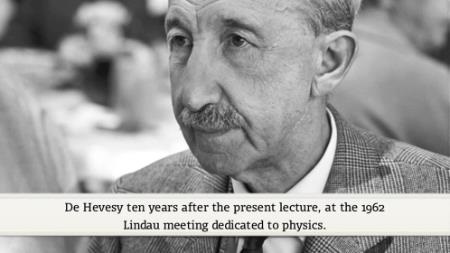
In 1952, de Hevesy participated in the second Lindau Meeting of Nobel Laureates, which was the first one dedicated to chemistry